The Critical Role of GMP-Grade UTP in mRNA Therapeutics and Biopharmaceutical Development

Introduction

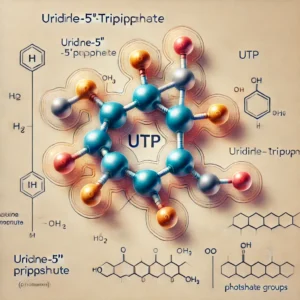
Uridine-5′-triphosphate (UTP) is a key nucleotide that plays a fundamental role in cellular metabolism, RNA synthesis, and signal transduction. In the rapidly evolving field of mRNA therapeutics, the use of high-quality, GMP (Good Manufacturing Practice)-grade UTP is critical to ensure the safety, efficacy, and regulatory compliance of pharmaceutical products. This article explores the significance of GMP-grade UTP, its applications in drug development, and the regulatory landscape governing its production and use.

Understanding Good Manufacturing Practice (GMP)

Good Manufacturing Practice (GMP) encompasses a set of stringent quality control guidelines established to ensure pharmaceutical products are consistently manufactured to the highest safety and efficacy standards. According to the World Health Organization (WHO), GMP ensures that products are “consistently produced and controlled according to quality standards appropriate for their intended use.” (WHO GMP Guidelines)

In the United States, the Food and Drug Administration (FDA) enforces current Good Manufacturing Practice (cGMP) regulations, which outline the minimum requirements for pharmaceutical manufacturers to ensure product quality. These regulations are designed to prevent contamination, ensure batch consistency, and guarantee that drugs meet rigorous quality standards. (FDA cGMP Overview)

Similarly, the European Medicines Agency (EMA) mandates that all pharmaceutical manufacturers supplying the European market must comply with GMP to maintain the highest standards of quality control. (EMA GMP Regulations)

The Role of GMP-Grade UTP in mRNA Therapeutics

mRNA-based therapies have gained immense traction, particularly in the development of vaccines and personalized medicine. The efficacy and safety of these therapies rely heavily on the purity and quality of raw materials, including nucleotides such as UTP.

  1. mRNA Synthesis
    • UTP is a crucial component in the in vitro transcription (IVT) process, where it serves as one of the four nucleotide triphosphates (NTPs) required for mRNA synthesis. The quality of UTP directly impacts the integrity, stability, and translation efficiency of the resulting mRNA molecule. (NIH Research on mRNA Synthesis)
  2. RNA Amplification
    • High-quality UTP is essential in RNA amplification technologies, ensuring accurate replication of genetic material for research and therapeutic purposes. (CDC RNA Amplification Guidelines)
  3. siRNA and Gene Editing Applications
    • In addition to mRNA vaccines, GMP-grade UTP is used in the production of small interfering RNA (siRNA) and gene-editing technologies such as CRISPR. The stringent quality control measures in GMP-certified production processes minimize risks of contamination and ensure reproducibility. (Genome.gov CRISPR Research)

Manufacturing and Quality Control of GMP-Grade UTP

GMP-grade UTP undergoes rigorous quality control assessments to meet regulatory standards and ensure product safety. The key aspects of its production include:

  • Purity and Testing:
    • GMP-grade UTP must exhibit high purity levels (>99%) as confirmed by High-Performance Liquid Chromatography (HPLC) and Mass Spectrometry (MS). (NIH Mass Spectrometry Standards)
  • Animal-Origin-Free (AOF) Production:
    • GMP-compliant manufacturers utilize animal-origin-free processes to mitigate risks associated with transmissible spongiform encephalopathies (TSE) and bovine spongiform encephalopathy (BSE). (FDA AOF Guidelines)
  • Sterility and Contamination Control:
  • Regulatory Documentation and Batch Traceability:
    • Comprehensive regulatory documentation supports compliance with FDA, EMA, and WHO requirements, ensuring traceability and accountability. (USP Pharmacopeial Standards)

Regulatory Considerations for GMP-Grade UTP

To maintain high manufacturing standards, regulatory agencies worldwide provide comprehensive guidelines for the production of nucleotides used in mRNA therapeutics:

  • FDA: The Biologics Control Act mandates strict oversight of biologics, including raw materials used in mRNA vaccines. (FDA Biologics Overview)
  • EMA: The Guideline on Quality, Non-Clinical, and Clinical Aspects of Gene Therapy Medicinal Products outlines stringent quality control measures. (EMA Gene Therapy Guidelines)
  • WHO: Provides global quality benchmarks for GMP-certified pharmaceutical ingredients. (WHO Pharmaceutical Standards)
Applications Beyond mRNA Therapeutics

Beyond its role in mRNA vaccine development, GMP-grade UTP is utilized in:

Conclusion

The demand for GMP-grade UTP continues to grow as mRNA-based technologies revolutionize medicine. Strict adherence to GMP standards ensures that UTP used in biopharmaceutical manufacturing is of the highest quality, minimizing risks and optimizing therapeutic outcomes. With regulatory agencies worldwide enforcing stringent quality measures, GMP-grade UTP remains an essential component in the advancement of mRNA therapeutics and beyond.

For further reading, visit WHO GMP Guidelines, FDA cGMP Regulations, and EMA GMP Guidelines.
 

The Advantages and Applications of Precast Protein Plus Gels in Modern Research

Introduction

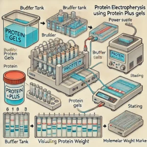
Electrophoresis is a fundamental technique in molecular biology and biochemistry, widely used for the separation and analysis of proteins. Traditionally, researchers had to prepare their own polyacrylamide gels, a labor-intensive process requiring precise formulation to achieve consistent results. The development of Precast Protein Plus Gels has revolutionized protein electrophoresis, providing scientists with a ready-to-use, high-performance alternative that enhances efficiency and reproducibility.

In this article, we explore the advantages, applications, and considerations of Precast Protein Plus Gels, including their impact on modern research and how they compare to traditional gel preparation. Additionally, we provide hyperlinks to trusted educational and governmental resources to further enhance understanding.

Advantages of Precast Protein Plus Gels

1. Time-Saving and User-Friendly

Traditional polyacrylamide gel preparation can be tedious and prone to inconsistencies. Precast Protein Plus Gels eliminate this step, allowing researchers to focus on data acquisition and analysis. These gels come pre-prepared and require no additional polymerization, reducing preparation time significantly (National Institutes of Health – NIH).

2. Consistency and Reproducibility

Manually cast gels often exhibit variability due to differences in polymerization conditions. Precast gels, manufactured under stringent quality controls, ensure uniform pore size and thickness, leading to reproducible results across multiple experiments (National Science Foundation – NSF).

3. Superior Resolution and Performance

High-quality Precast Protein Plus Gels provide sharper protein bands, which is critical for applications like Western blotting and proteomics. For instance, the Mini-PROTEAN TGX Gels by Bio-Rad offer excellent separation of polypeptides within a wide molecular weight range (Bio-Rad Laboratories).

4. Extended Shelf Life

Unlike manually prepared gels, which need immediate use, precast gels can be stored for months without degradation. Many brands offer shelf lives of up to 12 months when stored correctly (Food and Drug Administration – FDA).

5. Compatibility with Various Systems

Most Precast Protein Plus Gels are designed to fit common electrophoresis tanks, including those from major manufacturers such as Bio-Rad and Thermo Fisher (Thermo Fisher Scientific). This adaptability allows researchers to integrate them into existing workflows without additional equipment investments.

6. Reduced Risk of Contamination

Manual gel preparation involves multiple handling steps, increasing the risk of contamination. Precast gels minimize human intervention, reducing contamination and ensuring experimental accuracy (Centers for Disease Control and Prevention – CDC).

Applications of Precast Protein Plus Gels in Research

1. Protein Purity and Expression Analysis

Electrophoresis is essential for assessing protein purity in research and industrial applications. Precast Protein Plus Gels enable quick analysis of recombinant proteins used in drug development and structural biology (National Center for Biotechnology Information – NCBI).

2. Molecular Weight Estimation

Researchers rely on Precast Gels to estimate the molecular weight of proteins accurately. This application is crucial in characterizing unknown proteins and validating experimental findings (U.S. Department of Energy – DOE).

3. Western Blotting

Western blotting is a widely used technique to detect specific proteins in a sample. Precast Protein Plus Gels ensure superior resolution and compatibility with standard blotting protocols (National Institutes of Health – NIH).

4. Comparative Proteomics and Biomarker Discovery

Advancements in proteomics research demand high-throughput electrophoresis techniques. Precast gels are instrumental in comparing protein expression profiles, essential for biomarker discovery in diseases such as cancer and neurodegenerative disorders (National Cancer Institute – NCI).

5. Diagnostic Applications

Clinical laboratories use Precast Protein Plus Gels to analyze patient samples for protein-related diseases. This technique is crucial for diagnosing conditions like multiple myeloma and amyloidosis (World Health Organization – WHO).

Considerations for Selecting Precast Protein Plus Gels

1. Gel Percentage and Composition

The choice of gel percentage affects protein resolution. For example:

2. Buffer Systems

Different precast gels use distinct buffer systems. For example, Tris-Glycine gels are common, whereas Bis-Tris gels offer better stability and resolution at lower pH values (National Institute of Standards and Technology – NIST).

3. Well Formats and Sample Capacity

Precast gels come in various well configurations, including 10, 12, and 15-well formats. Choosing the right format depends on the number of samples to be analyzed (Environmental Protection Agency – EPA).

4. Compatibility with Staining and Detection Methods

Researchers using Precast Protein Plus Gels should ensure compatibility with staining techniques like Coomassie Blue, silver staining, and fluorescence-based detection (National Institute of Allergy and Infectious Diseases – NIAID).

Conclusion

Precast Protein Plus Gels have become indispensable in modern protein analysis due to their convenience, reproducibility, and superior performance. By eliminating manual preparation steps, these gels allow researchers to focus on obtaining high-quality results while minimizing variability and contamination risks. Their applications span from molecular weight estimation to biomarker discovery, making them a valuable tool in both academic and clinical research settings.

For further reading, refer to the authoritative resources linked throughout this article, which provide in-depth insights into electrophoresis techniques and protein analysis methodologies.

By integrating Precast Protein Plus Gels into their workflows, researchers can achieve greater efficiency and reliability, advancing scientific discoveries in proteomics, diagnostics, and biotechnology.


Annexin V-FITC/PI Apoptosis Detection Kit: A Reliable Assay for Apoptotic Cell Analysis

Introduction

The Annexin V-FITC/PI Apoptosis Detection Kit is a widely used tool for identifying apoptotic and necrotic cells. Apoptosis, or programmed cell death, plays a critical role in tissue homeostasis, immune responses, and disease pathogenesis. Detecting and quantifying apoptosis is essential in cancer research, drug discovery, and immunology studies.

This article explores the mechanism of apoptosis detection using the Annexin V-FITC/PI Kit, its applications, and protocol, with references to authoritative educational and government sources.

Understanding Apoptosis and Necrosis

Apoptosis is a tightly regulated cellular process, characterized by:

  • Phosphatidylserine (PS) Externalization: A hallmark of early apoptosis (NIH).
  • Chromatin Condensation and DNA Fragmentation: Features of late-stage apoptosis (NCBI).
  • Loss of Plasma Membrane Integrity: Indicative of necrosis rather than apoptosis (CDC).

How the Annexin V-FITC/PI Apoptosis Detection Kit Works

This kit employs two fluorescent probes:

  1. Annexin V-FITC (Fluorescein Isothiocyanate)
    • Binds to phosphatidylserine (PS) exposed on the outer leaflet of apoptotic cell membranes.
    • Detects early apoptotic cells (PubMed).
  2. Propidium Iodide (PI)
    • A DNA intercalating dye that stains necrotic and late-apoptotic cells with compromised membrane integrity.
    • Excludes viable and early apoptotic cells (National Cancer Institute).

Advantages of Annexin V-FITC/PI Assay

  • Highly Specific for Apoptosis: Differentiates between early and late apoptosis (FDA).
  • Rapid and Simple Protocol: Allows real-time apoptosis detection (Johns Hopkins Medicine).
  • Compatible with Flow Cytometry and Microscopy: Enables quantification and visualization (NIH Clinical Trials).
  • Non-toxic and Non-invasive: Preserves cell integrity for downstream assays (EPA).

Applications of Annexin V-FITC/PI Apoptosis Assay

1. Cancer Research

Apoptosis dysregulation is a hallmark of cancer, making this assay crucial for studying tumor cell death and therapeutic responses (National Cancer Institute).

2. Drug Screening and Toxicology

Assessing drug-induced apoptosis helps identify potential anti-cancer and cytotoxic compounds (FDA Research).

3. Immunology and Autoimmune Diseases

Understanding apoptosis in immune cells can provide insights into autoimmune disease mechanisms (NIH Immunology Research).

4. Neuroscience and Neurodegenerative Disorders

Apoptotic cell death is a key feature of neurodegenerative diseases like Alzheimer’s and Parkinson’s (National Institute of Neurological Disorders and Stroke).

Protocol for Annexin V-FITC/PI Assay

Step 1: Cell Preparation

  • Harvest adherent or suspension cells and wash with cold PBS (ATCC).
  • Resuspend cells in Annexin V binding buffer.

Step 2: Staining with Annexin V-FITC and PI

  • Add Annexin V-FITC and incubate for 15 minutes at room temperature in the dark.
  • Add PI solution to differentiate apoptotic and necrotic cells.

Step 3: Data Acquisition and Analysis

  • Analyze samples using flow cytometry or fluorescence microscopy.
  • Set up controls to distinguish live, early apoptotic, and necrotic cells (CDC Laboratory Training).

Interpretation of Results

  • Annexin V-FITC Positive / PI Negative: Early apoptosis.
  • Annexin V-FITC Positive / PI Positive: Late apoptosis or necrosis.
  • Annexin V-FITC Negative / PI Negative: Live cells.
  • Annexin V-FITC Negative / PI Positive: Necrotic cells (NIH Research).

Challenges and Considerations

  • Proper Controls: Include unstained, single-stained, and compensation controls for accurate gating.
  • Timing of Staining: Overstaining can lead to false positives.
  • Cell Concentration Optimization: Ensures accurate data interpretation (Mayo Clinic).

Future Directions in Apoptosis Detection

Innovations in multi-parametric flow cytometry, high-throughput screening, and AI-based image analysis are advancing apoptosis research. New biomarkers and live-cell imaging techniques will further enhance apoptosis detection (National Human Genome Research Institute).

Conclusion

The Annexin V-FITC/PI Apoptosis Detection Kit is a powerful tool for studying cell death, with applications in cancer research, toxicology, immunology, and neuroscience. Its ability to distinguish between live, apoptotic, and necrotic cells makes it indispensable for understanding disease mechanisms and therapeutic efficacy.


Rat Neuropilin-1 ELISA: A Key Tool for Neurological and Vascular Research

Introduction

Neuropilin-1 (NRP-1) is a multifunctional transmembrane glycoprotein that plays a crucial role in neuronal guidance, vascular development, and immune regulation. It is widely studied in neurological disorders, cancer biology, and cardiovascular research. The Rat Neuropilin-1 ELISA (Enzyme-Linked Immunosorbent Assay) is a highly sensitive method used to quantify NRP-1 levels in biological samples, such as serum, plasma, and tissue homogenates.

This article explores the significance of Neuropilin-1, the working principle of the ELISA method, its applications, and protocol steps, with references to authoritative educational and government sources.

Understanding Neuropilin-1 (NRP-1)

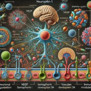
Neuropilin-1 is a co-receptor that interacts with multiple signaling pathways, particularly those involving vascular endothelial growth factor (VEGF) and semaphorin 3A. It plays a key role in:

How the Rat Neuropilin-1 ELISA Works

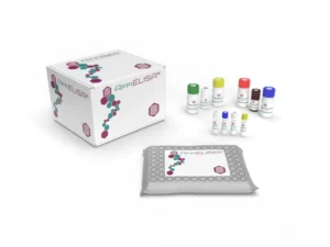
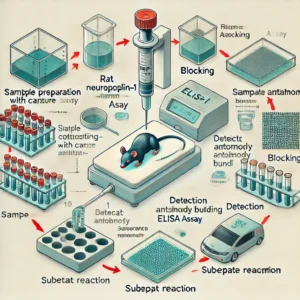
ELISA is a widely used immunoassay that relies on antigen-antibody interactions to detect and quantify proteins in biological fluids. The Rat Neuropilin-1 ELISA involves:

  1. Coating: Capture antibodies specific to NRP-1 are immobilized on a microplate (NCBI).
  2. Blocking: Non-specific binding sites are blocked to prevent background noise.
  3. Sample Incubation: Biological samples containing NRP-1 are added to the plate.
  4. Detection Antibody Binding: A biotinylated or enzyme-conjugated secondary antibody binds to NRP-1.
  5. Substrate Reaction: A colorimetric or chemiluminescent reaction generates a measurable signal (FDA).
  6. Quantification: Absorbance is measured at a specific wavelength to determine protein concentration (CDC).

Applications of Rat Neuropilin-1 ELISA

1. Neurological Research

NRP-1 is implicated in neurodevelopmental disorders, neurodegeneration, and axon guidance (Johns Hopkins Medicine).

2. Cancer Biology

NRP-1 is overexpressed in various cancers and is linked to tumor progression, metastasis, and angiogenesis (National Cancer Institute).

3. Cardiovascular Studies

NRP-1 regulates vascular permeability and endothelial function, making it a critical marker for cardiovascular diseases (American Heart Association).

4. Immune System and Autoimmunity

NRP-1 is involved in T-cell regulation and immune checkpoint pathways, making it a potential therapeutic target for autoimmune diseases (NIH Autoimmune Disease Research).

ELISA Protocol for Rat Neuropilin-1 Detection

Step 1: Sample Preparation

  • Collect serum, plasma, or tissue lysates from rat models.
  • Perform centrifugation to remove debris (ATCC).

Step 2: Plate Coating and Blocking

  • Coat a 96-well ELISA plate with capture antibody overnight at 4°C.
  • Block with a non-specific protein solution to reduce background signal.

Step 3: Sample and Antibody Incubation

  • Add biological samples to wells and incubate.
  • Introduce the detection antibody specific for NRP-1.

Step 4: Enzyme-Substrate Reaction

  • Add an enzyme-conjugated secondary antibody.
  • Introduce a substrate that reacts to generate a colorimetric signal (NIH Clinical Trials).

Step 5: Absorbance Measurement

  • Measure absorbance at 450 nm using a microplate reader (FDA Research).
  • Compare readings to a standard curve for quantification.

Interpretation of Results

  • High NRP-1 Levels: Indicate potential involvement in tumorigenesis, neuroinflammation, or cardiovascular disease.
  • Low NRP-1 Levels: Suggest normal physiological conditions or downregulated expression in disease models.
  • Standard Curve Calibration: Ensures precise quantification of unknown samples (CDC Laboratory Training).

Challenges and Considerations

  • Sample Quality: Proper sample handling and storage are crucial for reproducible results.
  • Assay Sensitivity: Optimization of antibody concentrations is required for specific detection.
  • Cross-reactivity: Use species-specific antibodies to avoid false positives (NIH Research).

Future Directions in Neuropilin-1 Research

Recent advancements in ELISA technology are improving the sensitivity and specificity of NRP-1 detection. Emerging techniques such as multiplex ELISA and nanotechnology-based biosensors offer promising avenues for enhancing biomarker discovery and therapeutic targeting (National Human Genome Research Institute).

Conclusion

The Rat Neuropilin-1 ELISA is a valuable tool for studying NRP-1 in neurological, oncological, and vascular research. Its high sensitivity and specificity make it an essential method for biomarker quantification. With ongoing advancements in ELISA technology, researchers can further refine their understanding of Neuropilin-1’s role in health and disease.


Ribo-off rRNA Depletion Kit: A Key Tool for RNA Sequencing and Transcriptomics

Introduction

The Ribo-off rRNA Depletion Kit is an essential tool for researchers conducting RNA sequencing (RNA-seq) and transcriptomic studies. This kit is designed to remove ribosomal RNA (rRNA) from total RNA samples, thereby enriching messenger RNA (mRNA) and long non-coding RNA (lncRNA) for more efficient and accurate downstream analysis. Ribosomal RNA makes up over 80% of total RNA in cells, and its depletion is crucial for obtaining high-quality sequencing data.

This article explores the importance of rRNA depletion, the mechanism of the Ribo-off rRNA Depletion Kit, its applications, and step-by-step protocol instructions, with references to authoritative educational and government sources.

Why rRNA Depletion is Important

Ribosomal RNA is highly abundant in total RNA extracts and can interfere with transcriptomic analysis. By removing rRNA, researchers can:

  • Improve sequencing depth for meaningful transcripts (NIH).
  • Reduce sequencing costs by focusing on biologically relevant RNA species (NCBI).
  • Enhance the accuracy of differential gene expression analysis (PubMed).
  • Increase the efficiency of library preparation and downstream bioinformatics analyses (National Cancer Institute).

How the Ribo-off rRNA Depletion Kit Works

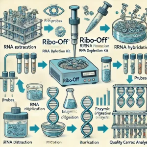
The Ribo-off rRNA Depletion Kit utilizes specifically designed probes that hybridize to rRNA molecules, allowing their selective removal. The workflow typically involves:

  1. rRNA Hybridization: rRNA-specific oligonucleotide probes bind to rRNA sequences.
  2. Enzymatic Digestion: Hybridized rRNA is degraded using RNAse enzymes.
  3. Cleanup and Purification: The remaining RNA, enriched in mRNA and lncRNA, is purified for further use (CDC).

Advantages of the Ribo-off rRNA Depletion Kit

  • Broad Compatibility: Works with different species, including human, mouse, and bacterial RNA (NCBI Gene Database).
  • High Efficiency: Removes up to 95% of rRNA, improving RNA-seq data quality (FDA).
  • Flexible Input: Compatible with low-input RNA samples for rare or limited biological materials (Johns Hopkins Medicine).
  • Cost-Effective: Reduces sequencing costs by prioritizing non-rRNA content (NIH Clinical Trials).

Applications of the Ribo-off rRNA Depletion Kit

1. RNA Sequencing (RNA-seq)

Depleting rRNA increases the efficiency of sequencing reads aligned to coding and non-coding RNAs, leading to better gene expression analysis (Mayo Clinic).

2. Transcriptomics and Gene Expression Studies

The removal of rRNA enables accurate profiling of lncRNA and small regulatory RNAs (NCATS).

3. Single-Cell RNA Sequencing

Enhances signal detection in single-cell transcriptomics by reducing rRNA-derived noise (NIH Stem Cell Information).

4. Microbiome and Metatranscriptomics Studies

Selective rRNA depletion helps researchers analyze microbial gene expression in environmental and medical microbiome research (EPA).

Protocol for rRNA Depletion Using Ribo-off

Step 1: RNA Preparation

Extract total RNA from cells or tissues using a high-quality RNA isolation kit (ATCC).

Step 2: Hybridization with rRNA Probes

Incubate the total RNA sample with rRNA depletion probes for optimal binding.

Step 3: Enzymatic Digestion

Add enzymatic digestion reagents to degrade rRNA molecules.

Step 4: Purification

Clean up the rRNA-depleted RNA using magnetic beads or column-based purification techniques (CDC Laboratory Training).

Step 5: Quality Control Check

Evaluate RNA integrity using a Bioanalyzer or TapeStation (National Human Genome Research Institute).

Interpretation of Results

  • High RNA Integrity Number (RIN): Indicates effective rRNA removal and sample quality.
  • Improved mRNA/LncRNA Yield: Demonstrates enrichment of biologically relevant RNA species.
  • Increased Data Usability: More meaningful sequencing data with reduced rRNA contamination.

Challenges and Considerations

  • Sample Quality: RNA integrity must be high before depletion to avoid degradation artifacts.
  • Species-Specific rRNA Removal: Ensure the correct probe set is used for the organism of interest.
  • Optimization for Low-Input Samples: Adjust conditions for working with minimal RNA amounts (FDA Research).

Future Directions in rRNA Depletion Technologies

Advancements in probe design, enzymatic efficiency, and automation are improving rRNA depletion methods. Emerging techniques such as CRISPR-based RNA depletion and machine-learning-driven transcriptomic analysis may further enhance RNA sequencing workflows (NIH Research).

Conclusion

The Ribo-off rRNA Depletion Kit is a powerful tool that enables researchers to improve the efficiency of RNA sequencing by removing rRNA contamination. Its applications extend across various fields, including transcriptomics, microbiome research, and precision medicine. By reducing sequencing costs and improving data accuracy, rRNA depletion is a critical step in modern molecular biology research.


Understanding Cell Counting Kit-8 (CCK-8) and Its Applications in Cell Viability Assays

Introduction

Cell Counting Kit-8 (CCK-8) is a widely used assay for measuring cell viability and proliferation. It is based on the reduction of a water-soluble tetrazolium salt (WST-8) by cellular dehydrogenases to produce a formazan dye, the intensity of which correlates with the number of viable cells. This non-radioactive, colorimetric assay is widely used in biomedical research, drug discovery, and toxicity testing.

This article provides a comprehensive overview of CCK-8, including its mechanism, advantages, applications, and protocol, with references to authoritative educational and government sources.

Mechanism of Action

The CCK-8 assay relies on the conversion of WST-8 into a highly water-soluble formazan product by intracellular dehydrogenases in viable cells. The formazan dye exhibits a strong absorbance at 450 nm, which can be quantitatively measured using a spectrophotometer (NCBI). This reaction occurs only in metabolically active cells, making it an accurate indicator of cell viability.

Advantages of CCK-8

  • High Sensitivity: Capable of detecting low numbers of viable cells (NIH).
  • Non-Toxicity: Unlike MTT assays, CCK-8 does not require cell lysis, allowing continuous monitoring of the same culture (CDC).
  • Water-Soluble Product: The formazan dye does not require solubilization, simplifying the assay workflow.
  • Reduced Background Interference: Improved stability and solubility result in higher signal-to-noise ratios (PubMed).
  • Compatibility: Suitable for high-throughput screening and robotic automation (NCATS).

Applications of CCK-8

1. Cell Proliferation Studies

CCK-8 is used to evaluate cell growth and division rates in response to external stimuli such as cytokines and growth factors (Johns Hopkins Medicine).

2. Drug Screening and Cytotoxicity Testing

Pharmaceutical researchers utilize CCK-8 to assess drug efficacy and toxicity in preclinical studies (FDA).

3. Cancer Research

CCK-8 aids in evaluating the effectiveness of anticancer drugs on tumor cells by measuring cell viability after treatment (National Cancer Institute).

4. Stem Cell Research

It is widely applied in stem cell studies to assess differentiation potential and cellular responses (NIH Stem Cell Information).

5. Toxicology and Environmental Studies

The assay is employed to study the cytotoxic effects of environmental pollutants and chemicals (EPA).

CCK-8 Protocol

  1. Cell Seeding: Plate cells in a 96-well plate and allow them to adhere overnight.
  2. Reagent Addition: Add 10 µL of CCK-8 solution per 100 µL of culture medium (ATCC).
  3. Incubation: Incubate for 1-4 hours at 37°C in a CO₂ incubator.
  4. Absorbance Measurement: Measure absorbance at 450 nm using a microplate reader (CDC Laboratory Training).
  5. Data Analysis: Compare absorbance values to control wells to determine cell viability.

Interpretation of Results

  • High Absorbance: Indicates a higher number of viable cells.
  • Low Absorbance: Suggests reduced cell viability or cytotoxicity.
  • Negative Control: Used to subtract background absorbance from media components (NIH Clinical Trials).

Challenges and Considerations

  • Assay Linearity: Requires optimization of cell density to ensure accurate results.
  • Interference from Test Compounds: Some drugs or natural extracts may interfere with WST-8 conversion, requiring parallel control experiments (Mayo Clinic).
  • Incubation Time: Over-incubation may lead to non-specific background signals.

Future Directions

Recent advancements in bioassays are improving the efficiency and accuracy of cell viability measurements. Integration with microfluidics and artificial intelligence-based image analysis is expected to enhance real-time monitoring of cell health in drug discovery (FDA Research).

Conclusion

Cell Counting Kit-8 (CCK-8) is an essential tool in cell biology and biomedical research. Its ease of use, sensitivity, and compatibility with various applications make it an invaluable assay for assessing cell viability and proliferation. Continued research and technological improvements will further refine its utility, enhancing biomedical advancements in cell-based studies.


Prenatal Risk Factors for Developmental Delay

When does pregnancy start?

The beginning of pregnancy is actually the first day of your last menstrual period. This is called gestational age or menstrual age. It is about two weeks before conception actually occurs. Although it may seem strange, the date of the first day of your last period will be an important date in determining your due date. Your health care provider will ask you about this date and use it to determine how far along you are in your pregnancy.

How does conception work?

Every month, your body goes through a reproductive cycle that can end in one of two ways. Either you will have a menstrual period or you will get pregnant. This cycle occurs continuously during your reproductive years, from puberty in your teens to menopause around age 50. In a cycle that ends with pregnancy, there are several steps. First, a group of eggs (called oocytes) prepare to leave the ovary for ovulation (egg release). The eggs develop in small fluid-filled cysts called follicles.

Think of these follicles as little containers for each immature egg. From this group of eggs, one will mature and continue through the cycle. This follicle then suppresses all other follicles in the group. The other follicles stop growing at this point. The mature follicle now breaks open and releases the egg from the ovary. This is ovulation. Ovulation usually occurs about two weeks before your next menstrual period starts. It is usually in the middle of its cycle.

After ovulation, the open (ruptured) follicle develops into a structure called the corpus luteum. It secretes (releases) the hormones progesterone and estrogen. Progesterone helps prepare the endometrium (lining of the uterus). This lining is where a fertilized egg settles to develop. If you don’t get pregnant during a cycle, this lining is what is shed during your period. On average, fertilization occurs about two weeks after your last menstrual period. When the sperm enters the egg, changes occur in the protein coat of the egg to prevent other sperm from entering.

At the time of fertilization, your baby’s genetic makeup is complete, including her gender. The sex of your baby depends on which sperm fertilizes the egg at the time of conception. Generally, women have a genetic combination of XX and men have XY. Women give each egg an X. Each sperm can be either an X or a Y. If the fertilized egg and sperm are a combination of an X and a Y, it’s a boy. If there are two X’s, it’s a girl.

What happens right after conception?

Within 24 hours after fertilization, the egg begins to rapidly divide into many cells. It stays in the fallopian tube for about three days after conception. The fertilized egg (now called a blastocyst) then continues to divide as it slowly passes through the fallopian tube into the uterus. Once there, its next job is to adhere to the endometrium. This is called implantation.

However, before implantation, the blastocyst breaks out of its protective shell. When the blastocyst comes into contact with the endometrium, the two exchange hormones to help the blastocyst attach. Some women notice spotting (light bleeding) for a day or two when implantation occurs. This is normal and not something to worry about.

At this point, the endometrium thickens and the cervix (the opening between the uterus and the birth canal) is sealed with a plug of mucus. In three weeks, the blastocyst cells finally form a small ball or embryo. At that time, the first nerve cells have formed. Your developing fetus has already gone through a few name changes in the first few weeks of pregnancy. Generally, it is called an embryo from conception to the eighth week of development. After the eighth week, it is called a fetus until it is born.

How early can I know that I am pregnant?

From the moment of conception, the hormone human chorionic gonadotropin (hCG) will be present in your blood. This hormone is created by the cells that make up the placenta (a food source for the growing fetus). It is also the hormone detected in a pregnancy test. Even though this hormone is there from the beginning, it takes time for it to develop within your body. It usually takes three to four weeks from the first day of your last period for hCG to raise enough to be detected by pregnancy tests.

When should I contact my health care provider about a new pregnancy?

Most health care providers will ask you to wait for an appointment until you have had a positive home pregnancy test. These tests are very accurate once you have enough hCG circulating throughout your body. This can be a few weeks after conception. It’s best to call your health care provider once you have a positive pregnancy test to schedule your first appointment.

When you call, your health care provider may ask if you are taking a prenatal vitamin. These supplements contain folic acid. It is important that you get at least 400 mcg of folic acid every day during pregnancy to ensure that the fetus’s neural tube (beginning of the brain and spinal column) develops properly. Many health care providers suggest that you take prenatal vitamins with folic acid even when you are not pregnant. If you weren’t taking prenatal vitamins before your pregnancy, your provider may ask you to start as soon as possible.

What is the timeline for fetal development?

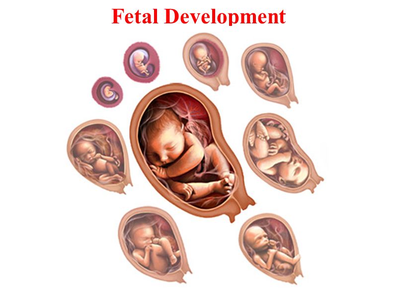
The fetus will change a lot during a typical pregnancy. This time is divided into three stages, called trimesters. Each trimester is a set of about three months. Your health care provider will probably talk to you about fetal development and risk in terms of weeks. So if you are three months pregnant, you are around 12 weeks.

You will see different changes in the fetus and yourself during each trimester.

Traditionally, we think of pregnancy as a nine-month process. However, this is not always the case. A full-term pregnancy is 40 weeks or 280 days. Depending on the months you are pregnant (some are shorter and some are longer) and the week you give birth, you could be pregnant for nine months or 10 months. This is completely normal and healthy.

Once you get closer to the end of your pregnancy, you may hear several category names as you go into labour. These labels divide the last weeks of pregnancy. They are also used to look for certain complications in newborns. Babies born at or before early-term may be at higher risk for breathing, hearing, or learning problems than babies born a few weeks later at full term. When looking at these labels, it is important to know how they are written. You may see the week first (38) and then you will see two numbers separated by a slash (6/7). This represents how many days you currently have in the gestational week. So if you see 38 6/7, it means you are on day 6 of your 38th week.

The last weeks of pregnancy are divided into the following groups:

Early term: 37 0/7 weeks to 38 6/7 weeks.
Full term: 39 0/7 weeks to 40 6/7 weeks.
Late-term: 41 0/7 weeks to 41 6/7 weeks.
Post-term: 42 0/7 weeks onwards.

Talk to your health care provider about any questions you may have about gestational age and due date.

Eukaryote vs Prokaryote

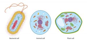
Every living organism falls into one of two groups: eukaryotes or prokaryotes. The cell structure determines which group an organism belongs to. In this article, we will explain in detail what prokaryotes and eukaryotes are and describe the differences between the two.

Definition of prokaryote

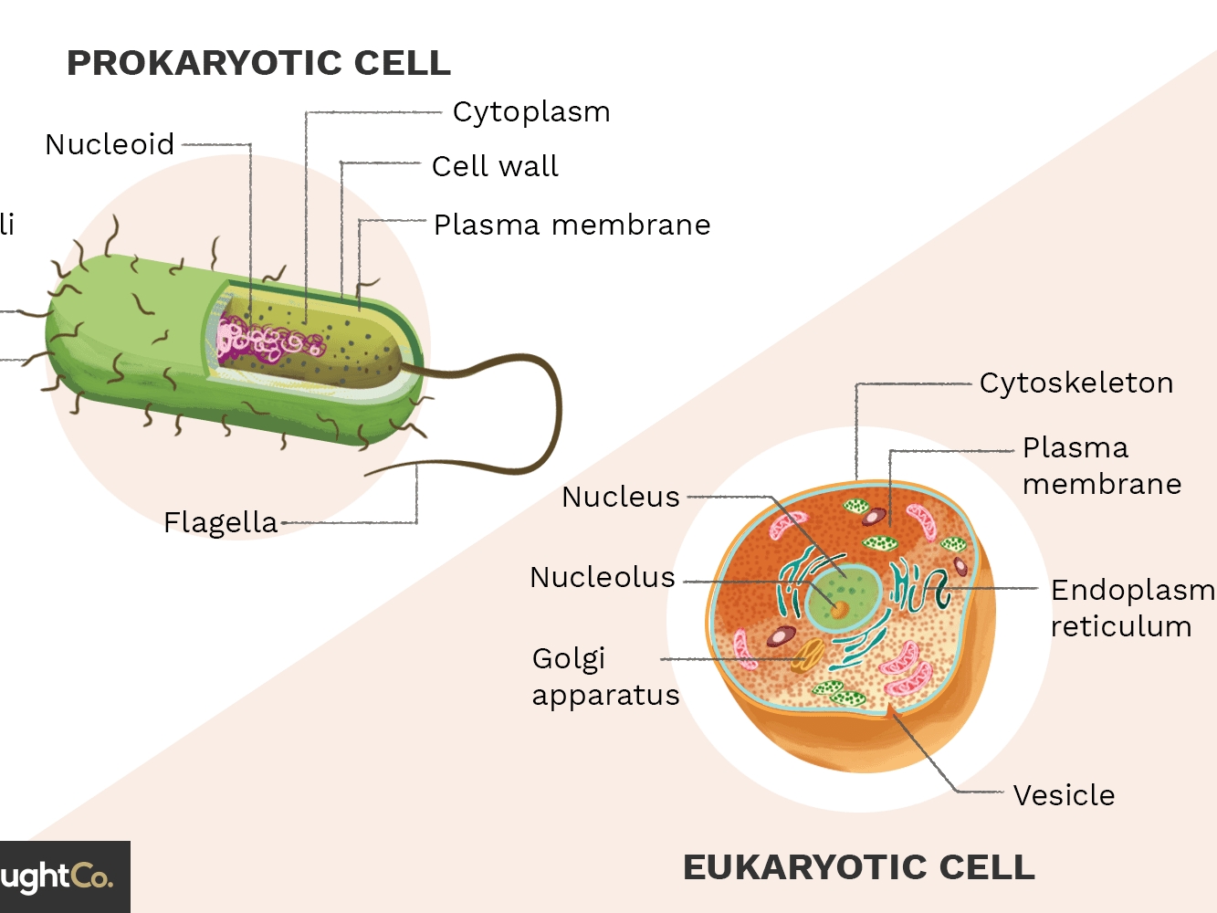
Prokaryotes are single-celled organisms that lack membrane-bound structures, the most notable of which is the nucleus. Prokaryotic cells tend to be small, simple cells, measuring between 0.1 and 5 μm in diameter. Although prokaryotic cells do not have membrane-bound structures, they do have distinct cellular regions. In prokaryotic cells, the DNA is grouped in a region called the nucleoid.

Characteristics of prokaryotic cells

Here’s a breakdown of what you might find in a prokaryotic bacterial cell.

  • Nucleoid: A central region of the cell that contains its DNA.
  • Ribosome: Ribosomes are responsible for protein synthesis.
  • Cell wall: The cell wall provides structure and protection from the outside environment. Most bacteria have a rigid cell wall made of carbohydrates and proteins called peptidoglycans.
  • Cell membrane: Every prokaryote has a cell membrane, also known as the plasma membrane, which separates the cell from the outside environment.
  • Capsule: Some bacteria have a layer of carbohydrates that surrounds the cell wall called a capsule. The capsule helps the bacteria stick to surfaces.
  • Fimbriae: Fimbriae are thin hair-like structures that help with cell attachment.
  • Pili: Pili are rod-shaped structures involved in multiple functions, including DNA binding and transfer.
  • Flagella: Flagella are thin, tail-like structures that aid in movement.

Examples of prokaryotes

Bacteria and archaea are the two types of prokaryotes.

Do prokaryotes have mitochondria?

No, prokaryotes do not have mitochondria. Mitochondria are only found in eukaryotic cells. This is also true for other membrane-bound structures, such as the nucleus and the Golgi apparatus (more on this later). One theory of eukaryotic evolution hypothesizes that mitochondria were the first prokaryotic cells to live inside other cells. Over time, evolution led these separate organisms to function as a single organism in the form of a eukaryote.

Definition of eukaryote

Eukaryotes are organisms whose cells have a nucleus and other organelles enclosed by a plasma membrane. Organelles are internal structures responsible for a variety of functions, such as energy production and protein synthesis. Eukaryotic cells are large (around 10-100 μm) and complex. While most eukaryotes are multicellular organisms, there are some single-celled eukaryotes.

Characteristics of eukaryotic cells

Within a eukaryotic cell, each membrane-bound structure carries out specific cellular functions. Here is an overview of many of the major components of eukaryotic cells.

  • Nucleus: The nucleus stores genetic information in the form of chromatin.
  • Nucleolus: Found within the nucleus, the nucleolus is the part of eukaryotic cells where ribosomal RNA is produced.
  • Plasma Membrane: The plasma membrane is a phospholipid bilayer that surrounds the entire cell and encompasses the internal organelles.
  • Cytoskeleton or cell wall: The cytoskeleton or cell wall provides structure, allows for cell movement, and plays a role in cell division.
  • Ribosomes: Ribosomes are responsible for protein synthesis.
  • Mitochondria: Mitochondria, also known as the power plants of the cell, are responsible for energy production.
  • Cytoplasm: The cytoplasm is the region of the cell between the nuclear envelope and the plasma membrane.
  • Cytosol: Cytosol is a gel-like substance inside the cell that contains the organelles.
  • Endoplasmic Reticulum: The endoplasmic reticulum is an organelle dedicated to protein maturation and transport.
  • Vesicles and vacuoles: Vesicles and vacuoles are membrane-bound sacs that are involved in transport and storage.

Other common organelles found in many, but not all, eukaryotes include the Golgi apparatus, chloroplasts, and lysosomes.

Examples of eukaryotes

Animals, plants, fungi, algae, and protozoa are all eukaryotes.

Comparing Prokaryotes and Eukaryotes

All life on Earth consists of eukaryotic cells or prokaryotic cells. Prokaryotes were the first life form. Scientists believe that eukaryotes evolved from prokaryotes about 2.7 billion years ago. The main distinction between these two types of organisms is that eukaryotic cells have a membrane-bound nucleus and prokaryotic cells do not. The nucleus is where eukaryotes store their genetic information.

In prokaryotes, DNA is bundled in the nucleoid region but is not stored within a membrane-bound nucleus. The nucleus is just one of many membrane-bound organelles in eukaryotes. Prokaryotes, on the other hand, do not have membrane-bound organelles. Another important difference is the structure of DNA. The DNA of eukaryotes consists of multiple linear double-stranded DNA molecules, while that of prokaryotes is double-stranded and circular.

Key Similarities Between Prokaryotes and Eukaryotes

All cells, whether prokaryotic or eukaryotic, share these four characteristics:

1. DNA

2. Plasma membrane

3. Cytoplasm

4. Ribosomes

Transcription and Translation in Prokaryotes vs. Eukaryotes

In prokaryotic cells, transcription and translation are coupled, meaning that translation begins during mRNA synthesis. In eukaryotic cells, transcription and translation are not coupled. Transcription occurs in the nucleus, producing mRNA. The mRNA then leaves the nucleus and translation occurs in the cytoplasm of the cell.

Endocytosis and Exocytosis

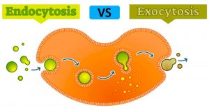
Endocytosis and exocytosis are the processes by which cells move materials into or out of the cell that is too large to pass directly through the lipid bilayer of the cell membrane. Large molecules, microorganisms, and waste products are some of the substances that move across the cell membrane through exocytosis and endocytosis.

Why is bulk transport important for cells?

Cell membranes are semi-permeable, meaning that they allow certain small molecules and ions to passively diffuse through them. Other small molecules can enter or leave the cell through carrier proteins or channels. But there are materials that are too large to pass through the cell membrane using these methods. There are times when a cell will need to engulf a bacterium or release a hormone. It is during these cases that bulk transport mechanisms are needed. Endocytosis and exocytosis are the bulk transport mechanisms used in eukaryotes. Since these transport processes require energy, they are known as active transport processes.

Vesicular function in endocytosis and exocytosis.

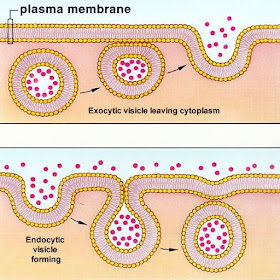
During bulk transport, larger substances or large packages of small molecules are transported across the cell membrane, also known as the plasma membrane, by means of vesicles; think of vesicles as little sacs of the membrane that can fuse with the cell membrane.

Cell membranes are composed of a lipid bilayer. The walls of the vesicles are also made up of a lipid bilayer, so they are capable of fusing with the cell membrane. This fusion between the vesicles and the plasma membrane facilitates bulk transport both in and out of the cell.

What is endocytosis? Endocytosis definition and purposes

Endocytosis is the process by which cells take in substances from outside the cell by engulfing them in a vesicle. These can include things like nutrients to support the cell or pathogens that the immune cells gobble up and destroy. Endocytosis occurs when a portion of the cell membrane folds back on itself, surrounding the extracellular fluid and various molecules or microorganisms. The resulting vesicle breaks apart and is transported into the cell.

Endocytosis serves many purposes, including:

  • Take in nutrients for cell growth, function, and repair: Cells need materials like proteins and lipids to function.
  • The capture of pathogens or other unknown substances that can endanger the body: When the immune system identifies pathogens such as bacteria, immune cells engulf them to destroy them.
  • Disposal of old or damaged cells: Cells must be disposed of safely when they stop working properly to prevent damage to other cells. These cells are removed by endocytosis.

Types of endocytosis

There are two types of endocytosis: phagocytosis and pinocytosis.

  • Phagocytosis

Phagocytosis, also known as cell ingestion, is the process by which cells internalize large cells or particles, such as damaged cells and bacteria. Within the human body and in other mammals, phagocytosis is the way immune cells engulf and destroy dangerous microorganisms or toxic compounds. Macrophages and neutrophils, types of white blood cells, are the two main phagocytes. These white blood cells are responsible for removing aged and damaged cells, as well as killing infectious microorganisms.

  • Pinocytosis

Pinocytosis, also known as cell drinking, is common in animal and plant cells. During pinocytosis, the cell takes up substances from the extracellular fluid that it needs to function. These include things like water and nutrients. Receptor-mediated endocytosis is a specialized type of pinocytosis. During receptor-mediated endocytosis, macromolecules bind to receptors along the surface of the cell’s plasma membrane. Cholesterol uptake is an example of receptor-mediated endocytosis.

The steps of endocytosis.

The following is a summary of the basic steps of the two types of endocytosis.

1. Phagocytosis:

  • A particle or substance binds to receptors on the cell surface, stimulating the release of pseudopods (cytoplasm-filled extensions of the plasma membrane).
  • The pseudopodia surround the object until their membranes fuse, forming a phagocytic vesicle.
  • The phagocytic vesicle detaches from the cell membrane and enters the cell.
  • The phagocytic vesicle fuses with lysosomes, which recycle or destroy the contents of the vesicle.

2. Pinocytosis:

  • The molecules bind to receptors located along the surface of the cell membrane.
  • The plasma membrane folds, forming a pinocytic vesicle that contains the molecules and extracellular fluid.
  • The pinocytic vesicle detaches from the cell membrane inside the cell.
  • The vesicle fuses with the first endosomes where the contents inside are sorted.

Example of endocytosis

Macrophages are a type of white blood cell that plays a central role in protecting mammals against pathogens such as bacteria and viruses. When a macrophage comes into contact with a virus, say a cold virus in the bloodstream, it can bind to the cell surface of the virus.

The macrophage will then form a vesicle around the virus, ingesting it completely. The vesicle then travels to the cytosol and fuses with the lysosome, where the virus is broken down. Some viruses replicate by “tricking” host cells into endocytosing them, at which point the virus hijacks the cell and tells it to replicate the virus genome and capsid.

What is exocytosis? Exocytosis definition and purposes

Exocytosis is the process by which cells move materials from inside the cell into the extracellular fluid. Exocytosis occurs when a vesicle fuses with the plasma membrane, allowing its contents to be released outside the cell.

Exocytosis has the following purposes:

  • Removal of toxins or waste products from within the cell: Cells create waste or toxins that must be removed from the cell to maintain homeostasis. For example, in aerobic respiration, cells produce the waste products carbon dioxide and water during the formation of ATP. Carbon dioxide and water are removed from these cells by exocytosis.
  • Facilitate cell communication: Cells create signalling molecules such as hormones and neurotransmitters. They are delivered to other cells upon their release from the cell through exocytosis.
  • Facilitate cell membrane growth, repair, signalling, and migration: When cells take in materials from outside the cell during endocytosis, they use lipids and proteins from the plasma membrane to create vesicles. When certain exocytotic vesicles fuse with the cell membrane, they replenish the cell membrane with these materials.

Types of exocytosis

  • Regulated exocytosis

Most exocytotic vesicles contain substances created within the endoplasmic reticulum for use elsewhere in the body, such as neurotransmitters or hormones. These molecules then pack inside a membrane layer called a vesicle. Once excreted from the endoplasmic reticulum, these vesicles are transported to the Golgi apparatus (also known as the Golgi complex) for further modification.

The molecules are then repackaged into a vesicle that makes its way to the plasma membrane. The release of these molecules from the cell is called regulated exocytosis because the expulsion of the materials is controlled or regulated by extracellular signals that cause membrane depolarization.

  • Constitutive exocytosis

Constitutive exocytosis, by contrast, does not require any extracellular signals. Most of the molecules that travel to the plasma membrane do so through this pathway.

After exocytosis, some exocytotic vesicles are incorporated into the plasma membrane (full vesicle fusion), while others return to the interior of the cell after their contents have been released (this is called the “kiss and run” pathway). Others remain attached to the membrane, where they can be used multiple times (the “kiss and stay” pathway).

The steps of exocytosis

Below is a summary of the basic steps of exocytosis.

  • A vesicle forms, typically within the endoplasmic reticulum and Golgi apparatus or early endosomes.
  • The vesicle travels to the cell membrane.
  • The vesicle fuses with the plasma membrane, during which the two bilayers fuse.
  • The contents of the vesicle are released into the extracellular space.
  • The vesicle fuses with or separates from the cell membrane.

Example of exocytosis

Let’s take the macrophage that we discussed in our endocytosis example. Once the white blood cell has engulfed a foreign pathogen, eliminate it, certain parts of the pathogen are no longer needed. The macrophage gets rid of this waste material through exocytosis, during which vesicles transport unwanted pathogenic material.

Development of antigen sandwich ELISA to detect interferon-alpha (IFN-α) using monoclonal antibodies in chicken

Development of antigen sandwich ELISA to detect interferon-alpha (IFN-α) using monoclonal antibodies in chicken

Interferon alpha (IFN-α) belongs to the kind I interferon household which mediates an early innate immune response to viral infections. Within the current examine, we developed sandwich ELISA utilizing particular mouse monoclonal antibodies (mAbs) to measure IFN-α manufacturing in chickens. Recombinant rooster IFN-α (chIFN-α) expressed in yeast have been bought from Kingfisher Biotech, and used to immunize the mice. 5 mAbs which particularly acknowledge rooster IFN-α antigen have been chosen and characterised. For sandwich ELISA improvement, mAbs have been labeled with biotin, adopted by a pairing take a look at to establish the perfect seize and detection antibodies. Two units of mouse anti-chIFN-α mAb pairs have been decided and an ordinary curve was established utilizing recombinant chIFN-α.

The sandwich ELISA successfully detected an elevated IFN-α manufacturing in rooster macrophage cells stimulated by polyinosinic:polycytidylic acid (poly I:C), and its minimal detectable stage was about 25 pg/mL. The anti-viral exercise of chIFN-α in opposition to vesicular stomatitis virus was characterised in avian embryonic fibroblast and the mouse anti-chIFN-α mAbs which neutralize its exercise have been recognized. The newly developed antigen sandwich ELISA developed on this examine will probably be a useful gizmo to watch IFN-α manufacturing in chickens.

The HIV Reservoirs Consortium: On this program, a consortium of educational labs has been established to outline the biology of the rebound-competent reservoir of HIV in vivo ( serological library) and, particularly, to find circulating non-viral biomarkers that can be utilized to watch it over time. A strategically centered, multidisciplinary workforce effort is finishing up research in PLHIV in resource-limited elements of the world in addition to in non-human primate fashions that recapitulate related points of human HIV an infection and during which the reservoir might be systematically perturbed with interventions that might not be utilized in people.

Utilizing state-of-the-art assays, it’s hoped that circulating non-viral biomarkers for the rebound-competent reservoir will probably be found within the non-human primate, cross-validated within the human, and assessed for his or her skill to outline the scale and high quality of the rebound-competent reservoir whereas on ART and the time to viral rebound as soon as ART is discontinued. Of notice, the HIV Frontiers Program pre-supposes the need to imagine and to share vital threat. Substantial new monetary sources and a sustained dedication will probably be required to pursue the a number of parts of a “single-shot” HIV remedy in parallel and to concurrently launch the HIV Reservoirs Consortium. Such sources and dedication is not going to come up from a single supply; quite, partnerships should be shaped and strategic priorities set. This assessment will define a number of the steps which might be being taken to achieve these objectives.

Development of antigen sandwich ELISA to detect interferon-alpha (IFN-α) using monoclonal antibodies in chicken

Modulating nonlinear elastic conduct of biodegradable form reminiscence elastomer and small intestinal submucosa(SIS) composites for smooth tissue restore

Structural restore of sentimental tissue for regenerative therapies might be superior by growing biocompatible and bioresorbable supplies with mechanical properties much like the tissue focused for remedy. Growing new supplies modeling smooth tissue mechanics can mitigate many limitations of fabric based mostly therapies, particularly regarding the mechanical stress and deformation the fabric imposes on surrounding tissue constructions. Nonetheless, many elastomeric supplies utilized in smooth tissue restore lack the power to be delivered by minimally invasive surgical (MIS) or transcatheter routes and require open surgical approaches for placement and utility.
We’ve developed a biocompatible and absolutely biodegradable form reminiscence elastomer, poly-(glycerol dodecanedioate) (PGD), which fulfills the necessities for hyperelasticity and reveals form reminiscence conduct to function a novel substrate materials for regenerative remedy in minimally invasive scientific procedures. Our earlier work demonstrated management over the tangent modulus at 12.5% compressive pressure between 1 and three MPa by rising the crosslinking density within the polymer. So as to enhance management over a broader vary of mechanical properties, nonlinear conduct, and toughness, we 1) diversified PGD bodily crosslink density, 2) included sheets of porcine small intestinal submucosa (SIS, Prepare dinner Biotech, Inc.) with various thickness, and three) combined lyophilized SIS particulates into PGD at completely different weight percentages.
Tensile testing (ASTM D412a) revealed PGD containing SIS sheets of have been stiffer than controls (p < 0.01). Incorporating lyophilized SIS particulates into PGD elevated the pressure to failure (p < 0.001) in comparison with PGD controls. Take a look at specimens with 1 ply sheets had higher tear power (ASTM D624c) in comparison with PGD tear specimens ready management specimens (p < 0.001). Nonetheless, incorporating SIS particulates decreased tear power of PGD-SIS 0.5 wt% particulate composites (p < 0.01) in comparison with PGD controls. Incorporating 2 ply and four ply sheets and 0.5 wt% particulates into PGD decreased the fixity and restoration of composite supplies in comparison with controls (p < 0.01). Nonlinear modeling of stress pressure curves underneath uniaxial pressure demonstrated tunability of PGD-SIS composite supplies to mannequin numerous nonlinear smooth tissues. These findings assist using form reminiscence PGD-SIS composite supplies in the direction of the design of implantable units for quite a lot of smooth tissue regeneration purposes by minimally invasive surgical procedure.
Isolation of antimicrobial-resistant microbes with Biocidal from ocular infections could also be changing into extra frequent. Infections attributable to these microbes might be troublesome to deal with and result in poor outcomes. Nonetheless, new therapies are being developed which can assist enhance scientific outcomes. This assessment examines current reviews on the isolation of antibiotic-resistant microbes from ocular infections.

AnaPrep 12 Instrument

Z1321001 1 unit
EUR 27916.8
Description: A fully automated magnetic bead-based nucleic acid extraction platform that uses preprogrammed protocols and can process up to 12 samples simultaneously. With the AnaPrep 12 Extractor you will have the option to choose and work with a wide range of sample and reagent volumes. This sturdy, realiable and user-friendly machine will save you both time and expenses while ensuring consistently high quality performance and nucleic acids for your downstream applications.

Instrument Covertile

SD6013 100 Pieces/Box Ask for price

Add-on Instrument

C55 -
EUR 432
Description: Single Instrument Add-on

Cell fusion instrument

E1659 1 Unit Ask for price

Nucleic Acid Instrument

Smart-32 1 unit/set Ask for price

Nucleic Acid Instrument

Swift-96 1 unit/set Ask for price

q-16 qPCR Instrument

Z-genesig-q16 each
EUR 9375

q-32 qPCR Instrument

Z-genesig-q32 each
EUR 18750

MyGo Pro qPCR Instrument

R32-Z-MyGo-Pro each
EUR 13750

AGS8830-8 PCR instrument

AGS8830 1 unit/set Ask for price

q-16 qPCR Instrument - RUO

R10016 each
EUR 9375

q-32 qPCR Instrument - RUO

R10032 each
EUR 18750

Lite Surgical Instrument Kit

SP0010-L 1 PC Ask for price

BioDrop Instrument Cover - EACH

SPE6052 EACH
EUR 81

MyGo Pro ESR qPCR Instrument

R32-Z-MyGo-Pro-ESR each
EUR 15000

1000 ug/mL Cadusafos in HPLC Acetonitrile Volume: 1mL**ANALYZED BY LCMS INSTRUMENTATION** - 1ML

LCS-4826 1ML
EUR 264.6

1000 ug/mL Maltodextrin in HPLC Grade Water Volume: 1mL Ampule**ANALYZED BY LC INSTRUMENTATION** - 1ML

LCS-5705 1ML
EUR 157.95

9600 Instrument PCR Detection kit

TRI-B32M1C 90T
EUR 957.6

9600 Instrument PCR Detection kit

TRI-B32M1E 60T
EUR 672.84

9600 Instrument PCR Detection Kit

TRI-B32S1O 48
EUR 453.6

9600 Instrument PCR Detection Kit

TRI-B32S1P 60
EUR 567

INSTRUMENT REST, HORIZONTAL SLOTTED

I556 1EA
EUR 19.7

BioDrop PC Instrument Cover - EACH

SPE6054 EACH
EUR 125.55

ICP-MS Instrument Check Standard 1 - 125ML

CL-ICS-1 125ML
EUR 461.7

Stereotaxic Instrument Calibration On-site Service-RWD Stereotaxic Instrument (Manual, Digital, Automated)

Service 18 1 Each Ask for price

1000 ug/mL Isoxaben in HPLC Acetonitrile Volume: 1mL Ampule**ANALYZED BY LCMS INSTRUMENTATION** - 1ML

LCS-5275 1ML
EUR 344.25

Instrument Check Standard 8 - 125ML

CALMIX8-100 125ML
EUR 249.75

MyGo Mini S qPCR Instrument - Red

R16-Z-MYGO-MINI-S-(RED) each
EUR 8118.75

Enzyme Instrument Cleaner conc - PK6

TRI2010 PK6
EUR 259.2

MyGo Mini S qPCR Instrument - Blue

R16-Z-MYGO-MINI-S-(BLUE) each
EUR 8118.75

MyGo Mini S qPCR Instrument - Pink

R16-Z-MYGO-MINI-S-(PINK) each
EUR 8118.75

1000 ug/mL Norflurazon in HPLC Acetonitrile Volume: 1mL Ampule**ANALYZED BY LCMS INSTRUMENTATION** - 1ML

LCS-2800-ACN 1ML
EUR 180.9

1000 ug/mL Acetamiprid in HPLC Acetonitrile Volume: 1mL Ampule**ANALYZED BY LCMS INSTRUMENTATION** - 1ML

LCS-4677-ACN 1ML
EUR 143.1

1000 ug/mL Pymetrozin in HPLC Acetonitrile Volume: 1mL Ampule**ANALYZED BY LCMS INSTRUMENTATION** - 1ML

LCS-4967 1ML
EUR 244.35

1000 ug/mL Tralkoxydim in HPLC Acetonitrile Volume: 1mL Ampule**ANALYZED BY LCMS INSTRUMENTATION** - 1ML

LCS-5470 1ML
EUR 264.6

1000 ug/mL Clethodium in HPLC Acetonitrile Volume: 1mL Ampule**ANALYZED BY LCMS INSTRUMENTATION** - 1ML

LCS-6083-ACN 1ML
EUR 155.25

MyGo Mini S qPCR Instrument - Green

R16-Z-MYGO-MINI-S-(GREEN) each
EUR 8118.75

Gradient gene amplification instrument

E1532 1 Unit Ask for price

Microsurgery Instrument Kit for Mice

SP0003-M 1 PC Ask for price

MyGo Mini S qPCR Instrument - Orange

R16-Z-MYGO-MINI-S-(ORANGE) each
EUR 8118.75

Isocratic gene amplification instrument

E1531 1 Unit Ask for price

Bone Surgery Instrument Kit for Mice

SP0006-M 1 PC Ask for price

1000 ug/mL Diflubenzuron in HPLC Acetonitrile Volume: 1mL Ampule**ANALYZED BY LCMS INSTRUMENTATION** - 1ML

LCS-1537-ACN 1ML
EUR 244.35

1000 ug/mL Dimethomorph in HPLC Acetonitrile Volume: 1mL Ampule**ANALYZED BY LCMS INSTRUMENTATION** - 1ML

LCS-3970-ACN 1ML
EUR 147.15

1000 ug/mL Dichlorprop-P in HPLC Acetonitrile Volume: 1mL Ampule**ANALYZED BY LCMS INSTRUMENTATION** - 1ML

LCS-6021 1ML
EUR 139.05

ICP-MS Instrument Calibration Standard 1 - 125ML

CL-CAL-1 125ML
EUR 511.65

ICP Instrument Check Standard 10 - 125ML

CALMIX10-100 125ML
EUR 256.5

ICP Instrument Check Standard 10 - 500ML

CALMIX10-500 500ML
EUR 433.35

ICP Instrument Check Standard 3 - 125ML

CALMIX3-100 125ML
EUR 309.15

ICP Instrument Check Standard 3 - 500ML

CALMIX3-500 500ML
EUR 498.15

ICP Instrument Check Standard 4 - 125ML

CALMIX4-100 125ML
EUR 324

ICP Instrument Check Standard 4 - 500ML

CALMIX4-500 500ML
EUR 527.85

ICP Instrument Check Standard 7 - 125ML

CALMIX7-100 125ML
EUR 348.3

ICP Instrument Check Standard 7 - 500ML

CALMIX7-500 500ML
EUR 602.1

ICP Instrument Check Standard 8 - 500ML

CALMIX8-500 500ML
EUR 413.1

General Surgery Instrument Kit for Rat

SP0009-R 1 PC Ask for price

1000 ug/mL Prothioconazole in HPLC AcetonitrileVolume: 1mL Ampule **ANALYZED BY LCMS INSTRUMENTATION** - 1ML

LCS-4904-ACN 1ML
EUR 167.4

1000 ug/mL Chlorpyrifos Oxon in HPLC AcetonitrileVolume: 1mL Ampule ***ANALYZED BY LCMS INSTRUMENTATION*** - 1ML

LCS-968 1ML
EUR 253.8

General Surgery Instrument Kit for Mouse

SP0009-M 1 PC Ask for price

General Surgery Instrument Kit for Rodents

SP0001-G 1 PC Ask for price

MCAO Surgical Instrument Kit for Rat & Mice

SP0011-B 1 PC Ask for price

1000 ug/mL Devrinol(Napropamide) in HPLC AcetonitrileVolume: 1mL Ampule **ANALYZED BY LCMS INSTRUMENTATION** - 1ML

LCS-1150-ACN 1ML
EUR 135

1000 ug/mL Prallethrin(mix of isomers)in HPLC AcetonitrileVolume: 1mL Ampule ***ANALYZED BY LCMS INSTRUMENTATION*** - 1ML

LCS-5783 1ML
EUR 253.8

1000 ug/mL Pyraflufen-ethyl in HPLC AcentonitrileVolume: 1mL Ampule **ANALYZED BY LCMS INSTRUMENTATION** - 1ML

LCS-6024-ACN 1ML
EUR 135

ICP-MS Instrument Check Standard 3 - 125ML

CL-ICS-3 125ML
EUR 253.8

ICP-MS Instrument Check Standard 4 - 125ML

CL-ICS-4 125ML
EUR 168.75

ICP-MS Instrument Check Standard 5 - 125ML

CL-ICS-5 125ML
EUR 187.65

Basic Surgical Instrument Kit for Rat & Mice

SP0010-B 1 PC Ask for price

Instrument Check Standard 1 for Ion Chromatography - 125ML

ICMIX1-100 125ML
EUR 236.25

Automatic Nucleic Acid Extraction Instrument(32)

VNP-32P 1
EUR 8000

Automatic Nucleic Acid Extraction Instrument(96)

VNP-96 1
EUR 12000

ICP-MS Instrument Calibration Standard 2 - 125ML

CL-CAL-2 125ML
EUR 546.75

SaMag Blood DNA Extraction kit For use with SaMag-12/24 instruments; extraction of genomic DNA from whole blood, peripheral blood mononuclear cells or buffy coat

SM001 48
EUR 187.48

MyGo Mini S qPCR Instrument - Grey Limited Edition

R16-Z-MYGO-MINIS(GREY) each
EUR 8118.75

Brain Microdialysis Surgery Instrument Kit for Rat

SP0004-R 1 PC Ask for price

Univeral Dissecting Surgery Instrument Kit for Rat

SP0007-R 1 PC Ask for price

Microsurgery Instrument Kit for Rat, Cat, Rabbit etc.

SP0003-R 1 PC Ask for price

Brain Microdialysis Surgery Instrument Kit for Mice

SP0004-M 1 PC Ask for price

Brain Microinjection Surgery Instrument Kit for Rat

SP0005-R 1 PC Ask for price

Univeral Dissecting Surgery Instrument Kit for Mice

SP0007-M 1 PC Ask for price

Bone Surgery Instrument Kit for Rat, Cat, Rabbit etc.

SP0006-R 1 PC Ask for price

Brain Microinjection Surgery Instrument Kit for Mice

SP0005-M 1 PC Ask for price

MiniVac Plastic Tubing Instrument to Bottle Lid - EACH

ARG1242 EACH
EUR 20.05

General Ophthalmic Surgery Instrument Kit for Rodents

SP0002-G 1 PC Ask for price

0.5ML CAPLESS TUBE COMPATIBLE WITH ABI-310 INSTRUMENT.

TUB-310-NC 500/pk
EUR 340.8
Description: PCR Plates & Tubes; PCR Tubes - Axygen

Spinal cord injury model Surgery Instrument Kit for Rat

SP0012-R 1 PC Ask for price

Instrument Check Standard 2 for Ion Chromatography - 125ML

ICMIX2-100 125ML
EUR 263.25

Instrument Check Standard 3 for Ion Chromatography - 125ML

ICMIX3-100 125ML
EUR 279.45

Instrument Check Standard 4 for Ion Chromatography - 125ML

ICMIX4-100 125ML
EUR 207.9

Instrument Check Standard 5 for Ion Chromatography - 125ML

ICMIX5-100 125ML
EUR 257.85

Instrument Check Standard 6 for Ion Chromatography - 125ML

ICMIX6-100 125ML
EUR 348.3

Univeral Dissecting Surgery Instrument Kit for Cat, Rabbit

SP0007-C 1 PC Ask for price

Spinal cord injury model Surgery Instrument Kit for Mouse

SP0012-M 1 PC Ask for price

Cardio-cerebral Perfusion Surgery Instrument Kit for Rat

SP0008-R 1 PC Ask for price

Nitrogen Blowing Instrument; Evaporation System; Pressure Blowing Concentrator

E0330 1 Unit Ask for price

Nitrogen Blowing Instrument; Evaporation System; Pressure Blowing Concentrator

E0331 1 Unit Ask for price

Nitrogen Blowing Instrument; Evaporation System; Pressure Blowing Concentrator

E0332 1 Unit Ask for price

Nitrogen Blowing Instrument; Evaporation System; Pressure Blowing Concentrator

E0333 1 Unit Ask for price

Nitrogen Blowing Instrument; Evaporation System; Pressure Blowing Concentrator

E0334 1 Unit Ask for price

Nitrogen Blowing Instrument; Evaporation System; Pressure Blowing Concentrator

E0335 1 Unit Ask for price

Nitrogen Blowing Instrument; Evaporation System; Pressure Blowing Concentrator

E0336 1 Unit Ask for price

Instrument Hardware Accessories Set (comprised of A2,3,4,5,6,7 and 10)

A-5041-A 1 Unit Ask for price

Laptop for Automated Stereotaxic Instrument (software included)

71000-LT 1 PC Ask for price

Cardio-cerebral Perfusion Surgery Instrument Kit for Mice

SP0008-M 1 PC Ask for price

96-channel fully automatic nucleic acid extraction instrument

QN-AUT-96 1 Unit Ask for price

16-channel fully automatic nucleic acid extraction instrument

QP-AUT-16 1 Unit Ask for price

24-channel fully automatic nucleic acid extraction instrument

QP-AUT-24 1 Unit Ask for price

32-channel fully automatic nucleic acid extraction instrument

QP-AUT-32 1 Unit Ask for price

1000 ug/mL 35-Diiodo-4-hydroxybenzonitrile (Ioxynil) Matrix:HPLCAcetonitrile Volume: 1mL Ampule *ANALYZED BY LCMS INSTRUMENTATION* - 1ML

LCS-1554-ACN 1ML
EUR 255.15

Pure Water Machine for Biochemical Instrument (15L/h, Heal Force)

E5050 1 Unit Ask for price

Pure Water Machine for Biochemical Instrument (30L/h, Heal Force)

E5051 1 Unit Ask for price

Pure Water Machine for Biochemical Instrument (100L/h, Small Vertical Machine, Heal Force)

E5055 1 Unit Ask for price

Pure Water Machine for Biochemical Instrument (200L/h, Small Vertical Machine, Heal Force)

E5056 1 Unit Ask for price

Pure Water Machine for Biochemical Instrument (100L/h, EDI, Small Vertical Machine, Heal Force)

E5053 1 Unit Ask for price

Pure Water Machine for Biochemical Instrument (150L/h, EDI, Small Vertical Machine, Heal Force)

E5054 1 Unit Ask for price

Myco-LumiTM Luminescent Mycoplasma Detection Kit for Low Sensitivity Instrument

C0297M 100 Tests Ask for price

Myco-LumiTM Luminescent Mycoplasma Detection Kit for Low Sensitivity Instrument

C0297S 20 Tests Ask for price

Myco-LumiTM Luminescent Mycoplasma Detection Kit for High Sensitivity Instrument

C0298M 100 Tests Ask for price

Myco-LumiTM Luminescent Mycoplasma Detection Kit for High Sensitivity Instrument

C0298S 20 Tests Ask for price

RS-485 line cable with extension plug for connection of an additional instrument

4819-PE 1 Piece Ask for price

On-site Installation and Operation Service Package for Automated Stereotaxic Instrument

Service 17 1 Each Ask for price

Pure Water Machine for Biochemical Instrument (70L/h, EDI, Small Vertical Machine, Heal Force)

E5052 1 Unit Ask for price

AnaPrep 48 Instrument, Automated Nucleic Acid Preparation System: including 1st year depot service and warranty.

Z2211001 1
EUR 20825

Myco-LumiTM Luminescent Mycoplasma Detection Kit for Low Sensitivity Instrument (Free Trial)

C0297FT 5 Tests Ask for price

AnaPrep 12 Dx Instrument, Automated Nucleic Acid Preparation System: including 1st year depot service and warranty.

Z2111001 1
EUR 20825

Sa2Res-SAMPLE TO RESULT Fully automated DNA/RNA extractor, PCR setup and qPCR amplification instrument

S2R-48 each Ask for price

PMF-AIR D'INSTRUMENT.-BL3-RLL090 AC Signs

271483 Roll of 505 Mark(s)
EUR 280.21
Description: Description Dutch: Leidingmerker - Air d'instrumentation; Description French: Marqueurs en rouleau continu, sans pictogramme - Air d'instrumentation

PMF-AIR D'INSTRUMENT.-BL4-RLL090 AC Signs

271484 Roll of 220 Mark(s)
EUR 280.21
Description: Description Dutch: Leidingmerker - Air d'instrumentation; Description French: Marqueurs en rouleau continu, sans pictogramme - Air d'instrumentation

EVrich™ Automatic Isolation Instruments

JOT-EV02-02-01 48 samples per run
EUR 24616.42

AIR D'INSTRUMENTATION 250X26CARD-T1-P8 AC Signs

N001999 Card of 4 Mark(s)
EUR 13.16
Description: Description Dutch: Leidingmerker - Air d'instrumentation; Description French: Marqueurs individuels sur carte avec flèches prédécoupées, sans pictogramme - Air d'instrumentation
As well as, an outline of the event of some new antibiotic therapies is given. The current literature concerning antibiotic use and resistance, isolation of antibiotic-resistant microbes from ocular infections and the event of potential new antibiotics that can be utilized to deal with these infections was reviewed. Ocular microbial infections are a worldwide public well being problem as they may end up in imaginative and prescient loss which compromises high quality of life. Roughly 70 per cent of ocular infections are attributable to micro organism together with Chlamydia trachomatis, Staphylococcus aureus, and Pseudomonas aeruginosa and fungi comparable to Candida albicans, Aspergillus spp. and Fusarium spp.