The Critical Role of GMP-Grade UTP in mRNA Therapeutics and Biopharmaceutical Development

Introduction

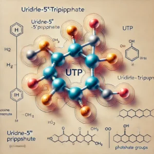
Uridine-5′-triphosphate (UTP) is a key nucleotide that plays a fundamental role in cellular metabolism, RNA synthesis, and signal transduction. In the rapidly evolving field of mRNA therapeutics, the use of high-quality, GMP (Good Manufacturing Practice)-grade UTP is critical to ensure the safety, efficacy, and regulatory compliance of pharmaceutical products. This article explores the significance of GMP-grade UTP, its applications in drug development, and the regulatory landscape governing its production and use.

Understanding Good Manufacturing Practice (GMP)

Good Manufacturing Practice (GMP) encompasses a set of stringent quality control guidelines established to ensure pharmaceutical products are consistently manufactured to the highest safety and efficacy standards. According to the World Health Organization (WHO), GMP ensures that products are “consistently produced and controlled according to quality standards appropriate for their intended use.” (WHO GMP Guidelines)

In the United States, the Food and Drug Administration (FDA) enforces current Good Manufacturing Practice (cGMP) regulations, which outline the minimum requirements for pharmaceutical manufacturers to ensure product quality. These regulations are designed to prevent contamination, ensure batch consistency, and guarantee that drugs meet rigorous quality standards. (FDA cGMP Overview)

Similarly, the European Medicines Agency (EMA) mandates that all pharmaceutical manufacturers supplying the European market must comply with GMP to maintain the highest standards of quality control. (EMA GMP Regulations)

The Role of GMP-Grade UTP in mRNA Therapeutics

mRNA-based therapies have gained immense traction, particularly in the development of vaccines and personalized medicine. The efficacy and safety of these therapies rely heavily on the purity and quality of raw materials, including nucleotides such as UTP.

  1. mRNA Synthesis
    • UTP is a crucial component in the in vitro transcription (IVT) process, where it serves as one of the four nucleotide triphosphates (NTPs) required for mRNA synthesis. The quality of UTP directly impacts the integrity, stability, and translation efficiency of the resulting mRNA molecule. (NIH Research on mRNA Synthesis)
  2. RNA Amplification
    • High-quality UTP is essential in RNA amplification technologies, ensuring accurate replication of genetic material for research and therapeutic purposes. (CDC RNA Amplification Guidelines)
  3. siRNA and Gene Editing Applications
    • In addition to mRNA vaccines, GMP-grade UTP is used in the production of small interfering RNA (siRNA) and gene-editing technologies such as CRISPR. The stringent quality control measures in GMP-certified production processes minimize risks of contamination and ensure reproducibility. (Genome.gov CRISPR Research)
Manufacturing and Quality Control of GMP-Grade UTP

GMP-grade UTP undergoes rigorous quality control assessments to meet regulatory standards and ensure product safety. The key aspects of its production include:

  • Purity and Testing:
    • GMP-grade UTP must exhibit high purity levels (>99%) as confirmed by High-Performance Liquid Chromatography (HPLC) and Mass Spectrometry (MS). (NIH Mass Spectrometry Standards)
  • Animal-Origin-Free (AOF) Production:
    • GMP-compliant manufacturers utilize animal-origin-free processes to mitigate risks associated with transmissible spongiform encephalopathies (TSE) and bovine spongiform encephalopathy (BSE). (FDA AOF Guidelines)
  • Sterility and Contamination Control:
  • Regulatory Documentation and Batch Traceability:
    • Comprehensive regulatory documentation supports compliance with FDA, EMA, and WHO requirements, ensuring traceability and accountability. (USP Pharmacopeial Standards)

Regulatory Considerations for GMP-Grade UTP

To maintain high manufacturing standards, regulatory agencies worldwide provide comprehensive guidelines for the production of nucleotides used in mRNA therapeutics:

  • FDA: The Biologics Control Act mandates strict oversight of biologics, including raw materials used in mRNA vaccines. (FDA Biologics Overview)
  • EMA: The Guideline on Quality, Non-Clinical, and Clinical Aspects of Gene Therapy Medicinal Products outlines stringent quality control measures. (EMA Gene Therapy Guidelines)
  • WHO: Provides global quality benchmarks for GMP-certified pharmaceutical ingredients. (WHO Pharmaceutical Standards)
Applications Beyond mRNA Therapeutics

Beyond its role in mRNA vaccine development, GMP-grade UTP is utilized in:

Conclusion

The demand for GMP-grade UTP continues to grow as mRNA-based technologies revolutionize medicine. Strict adherence to GMP standards ensures that UTP used in biopharmaceutical manufacturing is of the highest quality, minimizing risks and optimizing therapeutic outcomes. With regulatory agencies worldwide enforcing stringent quality measures, GMP-grade UTP remains an essential component in the advancement of mRNA therapeutics and beyond.

For further reading, visit WHO GMP Guidelines, FDA cGMP Regulations, and EMA GMP Guidelines.
 

The Advantages and Applications of Precast Protein Plus Gels in Modern Research

Introduction

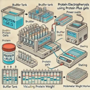
Electrophoresis is a fundamental technique in molecular biology and biochemistry, widely used for the separation and analysis of proteins. Traditionally, researchers had to prepare their own polyacrylamide gels, a labor-intensive process requiring precise formulation to achieve consistent results. The development of Precast Protein Plus Gels has revolutionized protein electrophoresis, providing scientists with a ready-to-use, high-performance alternative that enhances efficiency and reproducibility.

In this article, we explore the advantages, applications, and considerations of Precast Protein Plus Gels, including their impact on modern research and how they compare to traditional gel preparation. Additionally, we provide hyperlinks to trusted educational and governmental resources to further enhance understanding.

Advantages of Precast Protein Plus Gels

1. Time-Saving and User-Friendly

Traditional polyacrylamide gel preparation can be tedious and prone to inconsistencies. Precast Protein Plus Gels eliminate this step, allowing researchers to focus on data acquisition and analysis. These gels come pre-prepared and require no additional polymerization, reducing preparation time significantly (National Institutes of Health – NIH).

2. Consistency and Reproducibility

Manually cast gels often exhibit variability due to differences in polymerization conditions. Precast gels, manufactured under stringent quality controls, ensure uniform pore size and thickness, leading to reproducible results across multiple experiments (National Science Foundation – NSF).

3. Superior Resolution and Performance

High-quality Precast Protein Plus Gels provide sharper protein bands, which is critical for applications like Western blotting and proteomics. For instance, the Mini-PROTEAN TGX Gels by Bio-Rad offer excellent separation of polypeptides within a wide molecular weight range (Bio-Rad Laboratories).

4. Extended Shelf Life

Unlike manually prepared gels, which need immediate use, precast gels can be stored for months without degradation. Many brands offer shelf lives of up to 12 months when stored correctly (Food and Drug Administration – FDA).

5. Compatibility with Various Systems

Most Precast Protein Plus Gels are designed to fit common electrophoresis tanks, including those from major manufacturers such as Bio-Rad and Thermo Fisher (Thermo Fisher Scientific). This adaptability allows researchers to integrate them into existing workflows without additional equipment investments.

6. Reduced Risk of Contamination

Manual gel preparation involves multiple handling steps, increasing the risk of contamination. Precast gels minimize human intervention, reducing contamination and ensuring experimental accuracy (Centers for Disease Control and Prevention – CDC).

Applications of Precast Protein Plus Gels in Research

1. Protein Purity and Expression Analysis

Electrophoresis is essential for assessing protein purity in research and industrial applications. Precast Protein Plus Gels enable quick analysis of recombinant proteins used in drug development and structural biology (National Center for Biotechnology Information – NCBI).

2. Molecular Weight Estimation

Researchers rely on Precast Gels to estimate the molecular weight of proteins accurately. This application is crucial in characterizing unknown proteins and validating experimental findings (U.S. Department of Energy – DOE).

3. Western Blotting

Western blotting is a widely used technique to detect specific proteins in a sample. Precast Protein Plus Gels ensure superior resolution and compatibility with standard blotting protocols (National Institutes of Health – NIH).

4. Comparative Proteomics and Biomarker Discovery

Advancements in proteomics research demand high-throughput electrophoresis techniques. Precast gels are instrumental in comparing protein expression profiles, essential for biomarker discovery in diseases such as cancer and neurodegenerative disorders (National Cancer Institute – NCI).

5. Diagnostic Applications

Clinical laboratories use Precast Protein Plus Gels to analyze patient samples for protein-related diseases. This technique is crucial for diagnosing conditions like multiple myeloma and amyloidosis (World Health Organization – WHO).

Considerations for Selecting Precast Protein Plus Gels

1. Gel Percentage and Composition

The choice of gel percentage affects protein resolution. For example:

2. Buffer Systems

Different precast gels use distinct buffer systems. For example, Tris-Glycine gels are common, whereas Bis-Tris gels offer better stability and resolution at lower pH values (National Institute of Standards and Technology – NIST).

3. Well Formats and Sample Capacity

Precast gels come in various well configurations, including 10, 12, and 15-well formats. Choosing the right format depends on the number of samples to be analyzed (Environmental Protection Agency – EPA).

4. Compatibility with Staining and Detection Methods

Researchers using Precast Protein Plus Gels should ensure compatibility with staining techniques like Coomassie Blue, silver staining, and fluorescence-based detection (National Institute of Allergy and Infectious Diseases – NIAID).

Conclusion

Precast Protein Plus Gels have become indispensable in modern protein analysis due to their convenience, reproducibility, and superior performance. By eliminating manual preparation steps, these gels allow researchers to focus on obtaining high-quality results while minimizing variability and contamination risks. Their applications span from molecular weight estimation to biomarker discovery, making them a valuable tool in both academic and clinical research settings.

For further reading, refer to the authoritative resources linked throughout this article, which provide in-depth insights into electrophoresis techniques and protein analysis methodologies.

By integrating Precast Protein Plus Gels into their workflows, researchers can achieve greater efficiency and reliability, advancing scientific discoveries in proteomics, diagnostics, and biotechnology.


Annexin V-FITC/PI Apoptosis Detection Kit: A Reliable Assay for Apoptotic Cell Analysis

Introduction

The Annexin V-FITC/PI Apoptosis Detection Kit is a widely used tool for identifying apoptotic and necrotic cells. Apoptosis, or programmed cell death, plays a critical role in tissue homeostasis, immune responses, and disease pathogenesis. Detecting and quantifying apoptosis is essential in cancer research, drug discovery, and immunology studies.

This article explores the mechanism of apoptosis detection using the Annexin V-FITC/PI Kit, its applications, and protocol, with references to authoritative educational and government sources.

Understanding Apoptosis and Necrosis

Apoptosis is a tightly regulated cellular process, characterized by:

  • Phosphatidylserine (PS) Externalization: A hallmark of early apoptosis (NIH).
  • Chromatin Condensation and DNA Fragmentation: Features of late-stage apoptosis (NCBI).
  • Loss of Plasma Membrane Integrity: Indicative of necrosis rather than apoptosis (CDC).

How the Annexin V-FITC/PI Apoptosis Detection Kit Works

This kit employs two fluorescent probes:

  1. Annexin V-FITC (Fluorescein Isothiocyanate)
    • Binds to phosphatidylserine (PS) exposed on the outer leaflet of apoptotic cell membranes.
    • Detects early apoptotic cells (PubMed).
  2. Propidium Iodide (PI)
    • A DNA intercalating dye that stains necrotic and late-apoptotic cells with compromised membrane integrity.
    • Excludes viable and early apoptotic cells (National Cancer Institute).

Advantages of Annexin V-FITC/PI Assay

  • Highly Specific for Apoptosis: Differentiates between early and late apoptosis (FDA).
  • Rapid and Simple Protocol: Allows real-time apoptosis detection (Johns Hopkins Medicine).
  • Compatible with Flow Cytometry and Microscopy: Enables quantification and visualization (NIH Clinical Trials).
  • Non-toxic and Non-invasive: Preserves cell integrity for downstream assays (EPA).

Applications of Annexin V-FITC/PI Apoptosis Assay

1. Cancer Research

Apoptosis dysregulation is a hallmark of cancer, making this assay crucial for studying tumor cell death and therapeutic responses (National Cancer Institute).

2. Drug Screening and Toxicology

Assessing drug-induced apoptosis helps identify potential anti-cancer and cytotoxic compounds (FDA Research).

3. Immunology and Autoimmune Diseases

Understanding apoptosis in immune cells can provide insights into autoimmune disease mechanisms (NIH Immunology Research).

4. Neuroscience and Neurodegenerative Disorders

Apoptotic cell death is a key feature of neurodegenerative diseases like Alzheimer’s and Parkinson’s (National Institute of Neurological Disorders and Stroke).

Protocol for Annexin V-FITC/PI Assay

Step 1: Cell Preparation

  • Harvest adherent or suspension cells and wash with cold PBS (ATCC).
  • Resuspend cells in Annexin V binding buffer.

Step 2: Staining with Annexin V-FITC and PI

  • Add Annexin V-FITC and incubate for 15 minutes at room temperature in the dark.
  • Add PI solution to differentiate apoptotic and necrotic cells.

Step 3: Data Acquisition and Analysis

  • Analyze samples using flow cytometry or fluorescence microscopy.
  • Set up controls to distinguish live, early apoptotic, and necrotic cells (CDC Laboratory Training).

Interpretation of Results

  • Annexin V-FITC Positive / PI Negative: Early apoptosis.
  • Annexin V-FITC Positive / PI Positive: Late apoptosis or necrosis.
  • Annexin V-FITC Negative / PI Negative: Live cells.
  • Annexin V-FITC Negative / PI Positive: Necrotic cells (NIH Research).

Challenges and Considerations

  • Proper Controls: Include unstained, single-stained, and compensation controls for accurate gating.
  • Timing of Staining: Overstaining can lead to false positives.
  • Cell Concentration Optimization: Ensures accurate data interpretation (Mayo Clinic).

Future Directions in Apoptosis Detection

Innovations in multi-parametric flow cytometry, high-throughput screening, and AI-based image analysis are advancing apoptosis research. New biomarkers and live-cell imaging techniques will further enhance apoptosis detection (National Human Genome Research Institute).

Conclusion

The Annexin V-FITC/PI Apoptosis Detection Kit is a powerful tool for studying cell death, with applications in cancer research, toxicology, immunology, and neuroscience. Its ability to distinguish between live, apoptotic, and necrotic cells makes it indispensable for understanding disease mechanisms and therapeutic efficacy.


Rat Neuropilin-1 ELISA: A Key Tool for Neurological and Vascular Research

Introduction

Neuropilin-1 (NRP-1) is a multifunctional transmembrane glycoprotein that plays a crucial role in neuronal guidance, vascular development, and immune regulation. It is widely studied in neurological disorders, cancer biology, and cardiovascular research. The Rat Neuropilin-1 ELISA (Enzyme-Linked Immunosorbent Assay) is a highly sensitive method used to quantify NRP-1 levels in biological samples, such as serum, plasma, and tissue homogenates.

This article explores the significance of Neuropilin-1, the working principle of the ELISA method, its applications, and protocol steps, with references to authoritative educational and government sources.

Understanding Neuropilin-1 (NRP-1)

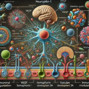
Neuropilin-1 is a co-receptor that interacts with multiple signaling pathways, particularly those involving vascular endothelial growth factor (VEGF) and semaphorin 3A. It plays a key role in:

How the Rat Neuropilin-1 ELISA Works

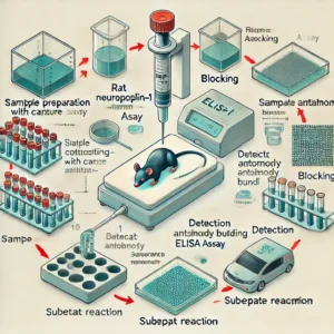
ELISA is a widely used immunoassay that relies on antigen-antibody interactions to detect and quantify proteins in biological fluids. The Rat Neuropilin-1 ELISA involves:

  1. Coating: Capture antibodies specific to NRP-1 are immobilized on a microplate (NCBI).
  2. Blocking: Non-specific binding sites are blocked to prevent background noise.
  3. Sample Incubation: Biological samples containing NRP-1 are added to the plate.
  4. Detection Antibody Binding: A biotinylated or enzyme-conjugated secondary antibody binds to NRP-1.
  5. Substrate Reaction: A colorimetric or chemiluminescent reaction generates a measurable signal (FDA).
  6. Quantification: Absorbance is measured at a specific wavelength to determine protein concentration (CDC).

Applications of Rat Neuropilin-1 ELISA

1. Neurological Research

NRP-1 is implicated in neurodevelopmental disorders, neurodegeneration, and axon guidance (Johns Hopkins Medicine).

2. Cancer Biology

NRP-1 is overexpressed in various cancers and is linked to tumor progression, metastasis, and angiogenesis (National Cancer Institute).

3. Cardiovascular Studies

NRP-1 regulates vascular permeability and endothelial function, making it a critical marker for cardiovascular diseases (American Heart Association).

4. Immune System and Autoimmunity

NRP-1 is involved in T-cell regulation and immune checkpoint pathways, making it a potential therapeutic target for autoimmune diseases (NIH Autoimmune Disease Research).

ELISA Protocol for Rat Neuropilin-1 Detection

Step 1: Sample Preparation

  • Collect serum, plasma, or tissue lysates from rat models.
  • Perform centrifugation to remove debris (ATCC).

Step 2: Plate Coating and Blocking

  • Coat a 96-well ELISA plate with capture antibody overnight at 4°C.
  • Block with a non-specific protein solution to reduce background signal.

Step 3: Sample and Antibody Incubation

  • Add biological samples to wells and incubate.
  • Introduce the detection antibody specific for NRP-1.

Step 4: Enzyme-Substrate Reaction

  • Add an enzyme-conjugated secondary antibody.
  • Introduce a substrate that reacts to generate a colorimetric signal (NIH Clinical Trials).

Step 5: Absorbance Measurement

  • Measure absorbance at 450 nm using a microplate reader (FDA Research).
  • Compare readings to a standard curve for quantification.

Interpretation of Results

  • High NRP-1 Levels: Indicate potential involvement in tumorigenesis, neuroinflammation, or cardiovascular disease.
  • Low NRP-1 Levels: Suggest normal physiological conditions or downregulated expression in disease models.
  • Standard Curve Calibration: Ensures precise quantification of unknown samples (CDC Laboratory Training).

Challenges and Considerations

  • Sample Quality: Proper sample handling and storage are crucial for reproducible results.
  • Assay Sensitivity: Optimization of antibody concentrations is required for specific detection.
  • Cross-reactivity: Use species-specific antibodies to avoid false positives (NIH Research).

Future Directions in Neuropilin-1 Research

Recent advancements in ELISA technology are improving the sensitivity and specificity of NRP-1 detection. Emerging techniques such as multiplex ELISA and nanotechnology-based biosensors offer promising avenues for enhancing biomarker discovery and therapeutic targeting (National Human Genome Research Institute).

Conclusion

The Rat Neuropilin-1 ELISA is a valuable tool for studying NRP-1 in neurological, oncological, and vascular research. Its high sensitivity and specificity make it an essential method for biomarker quantification. With ongoing advancements in ELISA technology, researchers can further refine their understanding of Neuropilin-1’s role in health and disease.


Ribo-off rRNA Depletion Kit: A Key Tool for RNA Sequencing and Transcriptomics

Introduction

The Ribo-off rRNA Depletion Kit is an essential tool for researchers conducting RNA sequencing (RNA-seq) and transcriptomic studies. This kit is designed to remove ribosomal RNA (rRNA) from total RNA samples, thereby enriching messenger RNA (mRNA) and long non-coding RNA (lncRNA) for more efficient and accurate downstream analysis. Ribosomal RNA makes up over 80% of total RNA in cells, and its depletion is crucial for obtaining high-quality sequencing data.

This article explores the importance of rRNA depletion, the mechanism of the Ribo-off rRNA Depletion Kit, its applications, and step-by-step protocol instructions, with references to authoritative educational and government sources.

Why rRNA Depletion is Important

Ribosomal RNA is highly abundant in total RNA extracts and can interfere with transcriptomic analysis. By removing rRNA, researchers can:

  • Improve sequencing depth for meaningful transcripts (NIH).
  • Reduce sequencing costs by focusing on biologically relevant RNA species (NCBI).
  • Enhance the accuracy of differential gene expression analysis (PubMed).
  • Increase the efficiency of library preparation and downstream bioinformatics analyses (National Cancer Institute).

How the Ribo-off rRNA Depletion Kit Works

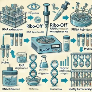
The Ribo-off rRNA Depletion Kit utilizes specifically designed probes that hybridize to rRNA molecules, allowing their selective removal. The workflow typically involves:

  1. rRNA Hybridization: rRNA-specific oligonucleotide probes bind to rRNA sequences.
  2. Enzymatic Digestion: Hybridized rRNA is degraded using RNAse enzymes.
  3. Cleanup and Purification: The remaining RNA, enriched in mRNA and lncRNA, is purified for further use (CDC).

Advantages of the Ribo-off rRNA Depletion Kit

  • Broad Compatibility: Works with different species, including human, mouse, and bacterial RNA (NCBI Gene Database).
  • High Efficiency: Removes up to 95% of rRNA, improving RNA-seq data quality (FDA).
  • Flexible Input: Compatible with low-input RNA samples for rare or limited biological materials (Johns Hopkins Medicine).
  • Cost-Effective: Reduces sequencing costs by prioritizing non-rRNA content (NIH Clinical Trials).

Applications of the Ribo-off rRNA Depletion Kit

1. RNA Sequencing (RNA-seq)

Depleting rRNA increases the efficiency of sequencing reads aligned to coding and non-coding RNAs, leading to better gene expression analysis (Mayo Clinic).

2. Transcriptomics and Gene Expression Studies

The removal of rRNA enables accurate profiling of lncRNA and small regulatory RNAs (NCATS).

3. Single-Cell RNA Sequencing

Enhances signal detection in single-cell transcriptomics by reducing rRNA-derived noise (NIH Stem Cell Information).

4. Microbiome and Metatranscriptomics Studies

Selective rRNA depletion helps researchers analyze microbial gene expression in environmental and medical microbiome research (EPA).

Protocol for rRNA Depletion Using Ribo-off

Step 1: RNA Preparation

Extract total RNA from cells or tissues using a high-quality RNA isolation kit (ATCC).

Step 2: Hybridization with rRNA Probes

Incubate the total RNA sample with rRNA depletion probes for optimal binding.

Step 3: Enzymatic Digestion

Add enzymatic digestion reagents to degrade rRNA molecules.

Step 4: Purification

Clean up the rRNA-depleted RNA using magnetic beads or column-based purification techniques (CDC Laboratory Training).

Step 5: Quality Control Check

Evaluate RNA integrity using a Bioanalyzer or TapeStation (National Human Genome Research Institute).

Interpretation of Results

  • High RNA Integrity Number (RIN): Indicates effective rRNA removal and sample quality.
  • Improved mRNA/LncRNA Yield: Demonstrates enrichment of biologically relevant RNA species.
  • Increased Data Usability: More meaningful sequencing data with reduced rRNA contamination.

Challenges and Considerations

  • Sample Quality: RNA integrity must be high before depletion to avoid degradation artifacts.
  • Species-Specific rRNA Removal: Ensure the correct probe set is used for the organism of interest.
  • Optimization for Low-Input Samples: Adjust conditions for working with minimal RNA amounts (FDA Research).

Future Directions in rRNA Depletion Technologies

Advancements in probe design, enzymatic efficiency, and automation are improving rRNA depletion methods. Emerging techniques such as CRISPR-based RNA depletion and machine-learning-driven transcriptomic analysis may further enhance RNA sequencing workflows (NIH Research).

Conclusion

The Ribo-off rRNA Depletion Kit is a powerful tool that enables researchers to improve the efficiency of RNA sequencing by removing rRNA contamination. Its applications extend across various fields, including transcriptomics, microbiome research, and precision medicine. By reducing sequencing costs and improving data accuracy, rRNA depletion is a critical step in modern molecular biology research.


Understanding Cell Counting Kit-8 (CCK-8) and Its Applications in Cell Viability Assays

Introduction

Cell Counting Kit-8 (CCK-8) is a widely used assay for measuring cell viability and proliferation. It is based on the reduction of a water-soluble tetrazolium salt (WST-8) by cellular dehydrogenases to produce a formazan dye, the intensity of which correlates with the number of viable cells. This non-radioactive, colorimetric assay is widely used in biomedical research, drug discovery, and toxicity testing.

This article provides a comprehensive overview of CCK-8, including its mechanism, advantages, applications, and protocol, with references to authoritative educational and government sources.

Mechanism of Action

The CCK-8 assay relies on the conversion of WST-8 into a highly water-soluble formazan product by intracellular dehydrogenases in viable cells. The formazan dye exhibits a strong absorbance at 450 nm, which can be quantitatively measured using a spectrophotometer (NCBI). This reaction occurs only in metabolically active cells, making it an accurate indicator of cell viability.

Advantages of CCK-8

  • High Sensitivity: Capable of detecting low numbers of viable cells (NIH).
  • Non-Toxicity: Unlike MTT assays, CCK-8 does not require cell lysis, allowing continuous monitoring of the same culture (CDC).
  • Water-Soluble Product: The formazan dye does not require solubilization, simplifying the assay workflow.
  • Reduced Background Interference: Improved stability and solubility result in higher signal-to-noise ratios (PubMed).
  • Compatibility: Suitable for high-throughput screening and robotic automation (NCATS).

Applications of CCK-8

1. Cell Proliferation Studies

CCK-8 is used to evaluate cell growth and division rates in response to external stimuli such as cytokines and growth factors (Johns Hopkins Medicine).

2. Drug Screening and Cytotoxicity Testing

Pharmaceutical researchers utilize CCK-8 to assess drug efficacy and toxicity in preclinical studies (FDA).

3. Cancer Research

CCK-8 aids in evaluating the effectiveness of anticancer drugs on tumor cells by measuring cell viability after treatment (National Cancer Institute).

4. Stem Cell Research

It is widely applied in stem cell studies to assess differentiation potential and cellular responses (NIH Stem Cell Information).

5. Toxicology and Environmental Studies

The assay is employed to study the cytotoxic effects of environmental pollutants and chemicals (EPA).

CCK-8 Protocol

  1. Cell Seeding: Plate cells in a 96-well plate and allow them to adhere overnight.
  2. Reagent Addition: Add 10 µL of CCK-8 solution per 100 µL of culture medium (ATCC).
  3. Incubation: Incubate for 1-4 hours at 37°C in a CO₂ incubator.
  4. Absorbance Measurement: Measure absorbance at 450 nm using a microplate reader (CDC Laboratory Training).
  5. Data Analysis: Compare absorbance values to control wells to determine cell viability.

Interpretation of Results

  • High Absorbance: Indicates a higher number of viable cells.
  • Low Absorbance: Suggests reduced cell viability or cytotoxicity.
  • Negative Control: Used to subtract background absorbance from media components (NIH Clinical Trials).

Challenges and Considerations

  • Assay Linearity: Requires optimization of cell density to ensure accurate results.
  • Interference from Test Compounds: Some drugs or natural extracts may interfere with WST-8 conversion, requiring parallel control experiments (Mayo Clinic).
  • Incubation Time: Over-incubation may lead to non-specific background signals.

Future Directions

Recent advancements in bioassays are improving the efficiency and accuracy of cell viability measurements. Integration with microfluidics and artificial intelligence-based image analysis is expected to enhance real-time monitoring of cell health in drug discovery (FDA Research).

Conclusion

Cell Counting Kit-8 (CCK-8) is an essential tool in cell biology and biomedical research. Its ease of use, sensitivity, and compatibility with various applications make it an invaluable assay for assessing cell viability and proliferation. Continued research and technological improvements will further refine its utility, enhancing biomedical advancements in cell-based studies.