Prenatal Risk Factors for Developmental Delay

When does pregnancy start?

The beginning of pregnancy is actually the first day of your last menstrual period. This is called gestational age or menstrual age. It is about two weeks before conception actually occurs. Although it may seem strange, the date of the first day of your last period will be an important date in determining your due date. Your health care provider will ask you about this date and use it to determine how far along you are in your pregnancy.

How does conception work?

Every month, your body goes through a reproductive cycle that can end in one of two ways. Either you will have a menstrual period or you will get pregnant. This cycle occurs continuously during your reproductive years, from puberty in your teens to menopause around age 50. In a cycle that ends with pregnancy, there are several steps. First, a group of eggs (called oocytes) prepare to leave the ovary for ovulation (egg release). The eggs develop in small fluid-filled cysts called follicles.

Think of these follicles as little containers for each immature egg. From this group of eggs, one will mature and continue through the cycle. This follicle then suppresses all other follicles in the group. The other follicles stop growing at this point. The mature follicle now breaks open and releases the egg from the ovary. This is ovulation. Ovulation usually occurs about two weeks before your next menstrual period starts. It is usually in the middle of its cycle.

After ovulation, the open (ruptured) follicle develops into a structure called the corpus luteum. It secretes (releases) the hormones progesterone and estrogen. Progesterone helps prepare the endometrium (lining of the uterus). This lining is where a fertilized egg settles to develop. If you don’t get pregnant during a cycle, this lining is what is shed during your period. On average, fertilization occurs about two weeks after your last menstrual period. When the sperm enters the egg, changes occur in the protein coat of the egg to prevent other sperm from entering.

At the time of fertilization, your baby’s genetic makeup is complete, including her gender. The sex of your baby depends on which sperm fertilizes the egg at the time of conception. Generally, women have a genetic combination of XX and men have XY. Women give each egg an X. Each sperm can be either an X or a Y. If the fertilized egg and sperm are a combination of an X and a Y, it’s a boy. If there are two X’s, it’s a girl.

What happens right after conception?

Within 24 hours after fertilization, the egg begins to rapidly divide into many cells. It stays in the fallopian tube for about three days after conception. The fertilized egg (now called a blastocyst) then continues to divide as it slowly passes through the fallopian tube into the uterus. Once there, its next job is to adhere to the endometrium. This is called implantation.

However, before implantation, the blastocyst breaks out of its protective shell. When the blastocyst comes into contact with the endometrium, the two exchange hormones to help the blastocyst attach. Some women notice spotting (light bleeding) for a day or two when implantation occurs. This is normal and not something to worry about.

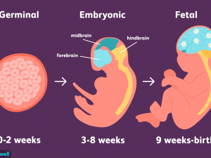
At this point, the endometrium thickens and the cervix (the opening between the uterus and the birth canal) is sealed with a plug of mucus. In three weeks, the blastocyst cells finally form a small ball or embryo. At that time, the first nerve cells have formed. Your developing fetus has already gone through a few name changes in the first few weeks of pregnancy. Generally, it is called an embryo from conception to the eighth week of development. After the eighth week, it is called a fetus until it is born.

How early can I know that I am pregnant?

From the moment of conception, the hormone human chorionic gonadotropin (hCG) will be present in your blood. This hormone is created by the cells that make up the placenta (a food source for the growing fetus). It is also the hormone detected in a pregnancy test. Even though this hormone is there from the beginning, it takes time for it to develop within your body. It usually takes three to four weeks from the first day of your last period for hCG to raise enough to be detected by pregnancy tests.

When should I contact my health care provider about a new pregnancy?

Most health care providers will ask you to wait for an appointment until you have had a positive home pregnancy test. These tests are very accurate once you have enough hCG circulating throughout your body. This can be a few weeks after conception. It’s best to call your health care provider once you have a positive pregnancy test to schedule your first appointment.

When you call, your health care provider may ask if you are taking a prenatal vitamin. These supplements contain folic acid. It is important that you get at least 400 mcg of folic acid every day during pregnancy to ensure that the fetus’s neural tube (beginning of the brain and spinal column) develops properly. Many health care providers suggest that you take prenatal vitamins with folic acid even when you are not pregnant. If you weren’t taking prenatal vitamins before your pregnancy, your provider may ask you to start as soon as possible.

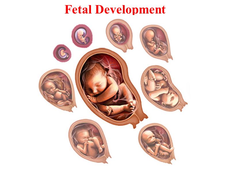
What is the timeline for fetal development?

The fetus will change a lot during a typical pregnancy. This time is divided into three stages, called trimesters. Each trimester is a set of about three months. Your health care provider will probably talk to you about fetal development and risk in terms of weeks. So if you are three months pregnant, you are around 12 weeks.

You will see different changes in the fetus and yourself during each trimester.

Traditionally, we think of pregnancy as a nine-month process. However, this is not always the case. A full-term pregnancy is 40 weeks or 280 days. Depending on the months you are pregnant (some are shorter and some are longer) and the week you give birth, you could be pregnant for nine months or 10 months. This is completely normal and healthy.

Once you get closer to the end of your pregnancy, you may hear several category names as you go into labour. These labels divide the last weeks of pregnancy. They are also used to look for certain complications in newborns. Babies born at or before early-term may be at higher risk for breathing, hearing, or learning problems than babies born a few weeks later at full term. When looking at these labels, it is important to know how they are written. You may see the week first (38) and then you will see two numbers separated by a slash (6/7). This represents how many days you currently have in the gestational week. So if you see 38 6/7, it means you are on day 6 of your 38th week.

The last weeks of pregnancy are divided into the following groups:

Early term: 37 0/7 weeks to 38 6/7 weeks.
Full term: 39 0/7 weeks to 40 6/7 weeks.
Late-term: 41 0/7 weeks to 41 6/7 weeks.
Post-term: 42 0/7 weeks onwards.

Talk to your health care provider about any questions you may have about gestational age and due date.

Eukaryote vs Prokaryote

Every living organism falls into one of two groups: eukaryotes or prokaryotes. The cell structure determines which group an organism belongs to. In this article, we will explain in detail what prokaryotes and eukaryotes are and describe the differences between the two.

Definition of prokaryote

Prokaryotes are single-celled organisms that lack membrane-bound structures, the most notable of which is the nucleus. Prokaryotic cells tend to be small, simple cells, measuring between 0.1 and 5 μm in diameter. Although prokaryotic cells do not have membrane-bound structures, they do have distinct cellular regions. In prokaryotic cells, the DNA is grouped in a region called the nucleoid.

Characteristics of prokaryotic cells

Here’s a breakdown of what you might find in a prokaryotic bacterial cell.

  • Nucleoid: A central region of the cell that contains its DNA.
  • Ribosome: Ribosomes are responsible for protein synthesis.
  • Cell wall: The cell wall provides structure and protection from the outside environment. Most bacteria have a rigid cell wall made of carbohydrates and proteins called peptidoglycans.
  • Cell membrane: Every prokaryote has a cell membrane, also known as the plasma membrane, which separates the cell from the outside environment.
  • Capsule: Some bacteria have a layer of carbohydrates that surrounds the cell wall called a capsule. The capsule helps the bacteria stick to surfaces.
  • Fimbriae: Fimbriae are thin hair-like structures that help with cell attachment.
  • Pili: Pili are rod-shaped structures involved in multiple functions, including DNA binding and transfer.
  • Flagella: Flagella are thin, tail-like structures that aid in movement.

Examples of prokaryotes

Bacteria and archaea are the two types of prokaryotes.

Do prokaryotes have mitochondria?

No, prokaryotes do not have mitochondria. Mitochondria are only found in eukaryotic cells. This is also true for other membrane-bound structures, such as the nucleus and the Golgi apparatus (more on this later). One theory of eukaryotic evolution hypothesizes that mitochondria were the first prokaryotic cells to live inside other cells. Over time, evolution led these separate organisms to function as a single organism in the form of a eukaryote.

Definition of eukaryote

Eukaryotes are organisms whose cells have a nucleus and other organelles enclosed by a plasma membrane. Organelles are internal structures responsible for a variety of functions, such as energy production and protein synthesis. Eukaryotic cells are large (around 10-100 μm) and complex. While most eukaryotes are multicellular organisms, there are some single-celled eukaryotes.

Characteristics of eukaryotic cells

Within a eukaryotic cell, each membrane-bound structure carries out specific cellular functions. Here is an overview of many of the major components of eukaryotic cells.

  • Nucleus: The nucleus stores genetic information in the form of chromatin.
  • Nucleolus: Found within the nucleus, the nucleolus is the part of eukaryotic cells where ribosomal RNA is produced.
  • Plasma Membrane: The plasma membrane is a phospholipid bilayer that surrounds the entire cell and encompasses the internal organelles.
  • Cytoskeleton or cell wall: The cytoskeleton or cell wall provides structure, allows for cell movement, and plays a role in cell division.
  • Ribosomes: Ribosomes are responsible for protein synthesis.
  • Mitochondria: Mitochondria, also known as the power plants of the cell, are responsible for energy production.
  • Cytoplasm: The cytoplasm is the region of the cell between the nuclear envelope and the plasma membrane.
  • Cytosol: Cytosol is a gel-like substance inside the cell that contains the organelles.
  • Endoplasmic Reticulum: The endoplasmic reticulum is an organelle dedicated to protein maturation and transport.
  • Vesicles and vacuoles: Vesicles and vacuoles are membrane-bound sacs that are involved in transport and storage.

Other common organelles found in many, but not all, eukaryotes include the Golgi apparatus, chloroplasts, and lysosomes.

Examples of eukaryotes

Animals, plants, fungi, algae, and protozoa are all eukaryotes.

Comparing Prokaryotes and Eukaryotes

All life on Earth consists of eukaryotic cells or prokaryotic cells. Prokaryotes were the first life form. Scientists believe that eukaryotes evolved from prokaryotes about 2.7 billion years ago. The main distinction between these two types of organisms is that eukaryotic cells have a membrane-bound nucleus and prokaryotic cells do not. The nucleus is where eukaryotes store their genetic information.

In prokaryotes, DNA is bundled in the nucleoid region but is not stored within a membrane-bound nucleus. The nucleus is just one of many membrane-bound organelles in eukaryotes. Prokaryotes, on the other hand, do not have membrane-bound organelles. Another important difference is the structure of DNA. The DNA of eukaryotes consists of multiple linear double-stranded DNA molecules, while that of prokaryotes is double-stranded and circular.

Key Similarities Between Prokaryotes and Eukaryotes

All cells, whether prokaryotic or eukaryotic, share these four characteristics:

1. DNA

2. Plasma membrane

3. Cytoplasm

4. Ribosomes

Transcription and Translation in Prokaryotes vs. Eukaryotes

In prokaryotic cells, transcription and translation are coupled, meaning that translation begins during mRNA synthesis. In eukaryotic cells, transcription and translation are not coupled. Transcription occurs in the nucleus, producing mRNA. The mRNA then leaves the nucleus and translation occurs in the cytoplasm of the cell.

Endocytosis and Exocytosis

Endocytosis and exocytosis are the processes by which cells move materials into or out of the cell that is too large to pass directly through the lipid bilayer of the cell membrane. Large molecules, microorganisms, and waste products are some of the substances that move across the cell membrane through exocytosis and endocytosis.

Why is bulk transport important for cells?

Cell membranes are semi-permeable, meaning that they allow certain small molecules and ions to passively diffuse through them. Other small molecules can enter or leave the cell through carrier proteins or channels. But there are materials that are too large to pass through the cell membrane using these methods. There are times when a cell will need to engulf a bacterium or release a hormone. It is during these cases that bulk transport mechanisms are needed. Endocytosis and exocytosis are the bulk transport mechanisms used in eukaryotes. Since these transport processes require energy, they are known as active transport processes.

Vesicular function in endocytosis and exocytosis.

During bulk transport, larger substances or large packages of small molecules are transported across the cell membrane, also known as the plasma membrane, by means of vesicles; think of vesicles as little sacs of the membrane that can fuse with the cell membrane.

Cell membranes are composed of a lipid bilayer. The walls of the vesicles are also made up of a lipid bilayer, so they are capable of fusing with the cell membrane. This fusion between the vesicles and the plasma membrane facilitates bulk transport both in and out of the cell.

What is endocytosis? Endocytosis definition and purposes

Endocytosis is the process by which cells take in substances from outside the cell by engulfing them in a vesicle. These can include things like nutrients to support the cell or pathogens that the immune cells gobble up and destroy. Endocytosis occurs when a portion of the cell membrane folds back on itself, surrounding the extracellular fluid and various molecules or microorganisms. The resulting vesicle breaks apart and is transported into the cell.

Endocytosis serves many purposes, including:

  • Take in nutrients for cell growth, function, and repair: Cells need materials like proteins and lipids to function.
  • The capture of pathogens or other unknown substances that can endanger the body: When the immune system identifies pathogens such as bacteria, immune cells engulf them to destroy them.
  • Disposal of old or damaged cells: Cells must be disposed of safely when they stop working properly to prevent damage to other cells. These cells are removed by endocytosis.

Types of endocytosis

There are two types of endocytosis: phagocytosis and pinocytosis.

  • Phagocytosis

Phagocytosis, also known as cell ingestion, is the process by which cells internalize large cells or particles, such as damaged cells and bacteria. Within the human body and in other mammals, phagocytosis is the way immune cells engulf and destroy dangerous microorganisms or toxic compounds. Macrophages and neutrophils, types of white blood cells, are the two main phagocytes. These white blood cells are responsible for removing aged and damaged cells, as well as killing infectious microorganisms.

  • Pinocytosis

Pinocytosis, also known as cell drinking, is common in animal and plant cells. During pinocytosis, the cell takes up substances from the extracellular fluid that it needs to function. These include things like water and nutrients. Receptor-mediated endocytosis is a specialized type of pinocytosis. During receptor-mediated endocytosis, macromolecules bind to receptors along the surface of the cell’s plasma membrane. Cholesterol uptake is an example of receptor-mediated endocytosis.

The steps of endocytosis.

The following is a summary of the basic steps of the two types of endocytosis.

1. Phagocytosis:

  • A particle or substance binds to receptors on the cell surface, stimulating the release of pseudopods (cytoplasm-filled extensions of the plasma membrane).
  • The pseudopodia surround the object until their membranes fuse, forming a phagocytic vesicle.
  • The phagocytic vesicle detaches from the cell membrane and enters the cell.
  • The phagocytic vesicle fuses with lysosomes, which recycle or destroy the contents of the vesicle.

2. Pinocytosis:

  • The molecules bind to receptors located along the surface of the cell membrane.
  • The plasma membrane folds, forming a pinocytic vesicle that contains the molecules and extracellular fluid.
  • The pinocytic vesicle detaches from the cell membrane inside the cell.
  • The vesicle fuses with the first endosomes where the contents inside are sorted.

Example of endocytosis

Macrophages are a type of white blood cell that plays a central role in protecting mammals against pathogens such as bacteria and viruses. When a macrophage comes into contact with a virus, say a cold virus in the bloodstream, it can bind to the cell surface of the virus.

The macrophage will then form a vesicle around the virus, ingesting it completely. The vesicle then travels to the cytosol and fuses with the lysosome, where the virus is broken down. Some viruses replicate by “tricking” host cells into endocytosing them, at which point the virus hijacks the cell and tells it to replicate the virus genome and capsid.

What is exocytosis? Exocytosis definition and purposes

Exocytosis is the process by which cells move materials from inside the cell into the extracellular fluid. Exocytosis occurs when a vesicle fuses with the plasma membrane, allowing its contents to be released outside the cell.

Exocytosis has the following purposes:

  • Removal of toxins or waste products from within the cell: Cells create waste or toxins that must be removed from the cell to maintain homeostasis. For example, in aerobic respiration, cells produce the waste products carbon dioxide and water during the formation of ATP. Carbon dioxide and water are removed from these cells by exocytosis.
  • Facilitate cell communication: Cells create signalling molecules such as hormones and neurotransmitters. They are delivered to other cells upon their release from the cell through exocytosis.
  • Facilitate cell membrane growth, repair, signalling, and migration: When cells take in materials from outside the cell during endocytosis, they use lipids and proteins from the plasma membrane to create vesicles. When certain exocytotic vesicles fuse with the cell membrane, they replenish the cell membrane with these materials.

Types of exocytosis

  • Regulated exocytosis

Most exocytotic vesicles contain substances created within the endoplasmic reticulum for use elsewhere in the body, such as neurotransmitters or hormones. These molecules then pack inside a membrane layer called a vesicle. Once excreted from the endoplasmic reticulum, these vesicles are transported to the Golgi apparatus (also known as the Golgi complex) for further modification.

The molecules are then repackaged into a vesicle that makes its way to the plasma membrane. The release of these molecules from the cell is called regulated exocytosis because the expulsion of the materials is controlled or regulated by extracellular signals that cause membrane depolarization.

  • Constitutive exocytosis

Constitutive exocytosis, by contrast, does not require any extracellular signals. Most of the molecules that travel to the plasma membrane do so through this pathway.

After exocytosis, some exocytotic vesicles are incorporated into the plasma membrane (full vesicle fusion), while others return to the interior of the cell after their contents have been released (this is called the “kiss and run” pathway). Others remain attached to the membrane, where they can be used multiple times (the “kiss and stay” pathway).

The steps of exocytosis

Below is a summary of the basic steps of exocytosis.

  • A vesicle forms, typically within the endoplasmic reticulum and Golgi apparatus or early endosomes.
  • The vesicle travels to the cell membrane.
  • The vesicle fuses with the plasma membrane, during which the two bilayers fuse.
  • The contents of the vesicle are released into the extracellular space.
  • The vesicle fuses with or separates from the cell membrane.

Example of exocytosis

Let’s take the macrophage that we discussed in our endocytosis example. Once the white blood cell has engulfed a foreign pathogen, eliminate it, certain parts of the pathogen are no longer needed. The macrophage gets rid of this waste material through exocytosis, during which vesicles transport unwanted pathogenic material.